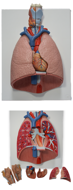
Main Model
C TRACHEA : 8 Pulmonary artery
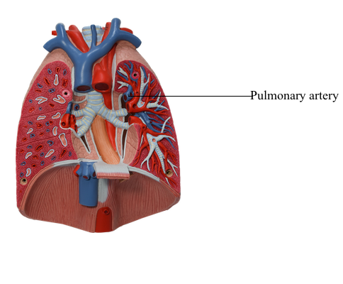
Each lung has a pulmonary artery supplying blood to it and
two pulmonary veins draining blood from it. The
right and left pulmonary arteries arise from the pulmonary trunk at the level of the sternal angle; they carry low-oxygen
("venous") blood to the lungs for oxygenation. (They are usually colored blue, like veins, or bluish-purple in anatomical
illustrations.) Each pulmonary artery becomes part of the root
of the corresponding lung and divides secondary lobar arteries. The right and left superior lobar arteries to the superior
lobes arise first, before entering the hilum. Continuing into
the lung, the artery descends posterolateral to the main bronchus as the inferior lobar artery of the left lung and as an intermediate artery that will divide into middle and inferior lobar
arteries of the right lung. Lobar arteries divide into tertiary segmental arteries. The arteries and bronchi are paired
in the lung, branching simultaneously and running parallel
courses. Consequently, a paired secondary lobar artery and
bronchus serves each lobe. Likewise, a paired tertiary segmental artery and bronchus supply each bronchopulmonary segment of the lung. Usually, the artery is located on the anterior aspect of the corresponding bronchus.
Two pulmonary veins, a superior and an inferior pulmonary vein on each side, carry oxygen-rich ("arterial") blood
from corresponding lobes of each lung to the left atrium of
the heart. The middle lobe vein is a tributary of the right
superior pulmonary vein. (Pulmonary veins are commonly
colored red, like arteries, or reddish-purple in anatomical
illustrations.) The pulmonary veins run independently of
the arteries and bronchi in the lung, coursing between and
receiving blood from adjacent bronchopulmonary segments as they run toward the hilum. Except in the central, perihilar
region of the lung, the veins from the visceral pleura and the bronchial venous circulation drain into the pulmonary veins,
the relatively small volume of low-oxygen blood entering the
large volume of oxygen-rich blood returning to the heart.
Veins from the parietal pleura join systemic veins in adjacent
parts of the thoracic wall.
Bronchial arteries supply blood for nutrition of the
structures making up the root of the lungs, the supporting tissues of the lungs, and the visceral pleura. The
two left bronchial arteries usually arise directly from the
thoracic aorta. The single right bronchial artery may also
arise directly from the aorta; however it commonly arises
indirectly, either by way of the proximal part of one of the
upper posterior intercostal arteries (usually the right 3rd posterior intercostal artery), or from a common trunk with the
left superior bronchial artery.
The small bronchial arteries provide branches to the
upper esophagus. Then they typically pass along the posterior aspects of the main bronchi, supplying them and their
branches as far distally as the respiratory bronchioles. The most distal branches of the
bronchial arteries anastomose with branches of the pulmonary arteries in the walls of the bronchioles and in the visceral pleura. The parietal pleura is supplied by the arteries
that supply the thoracic wall.
The bronchial veins drain only part of the
blood supplied to the lungs by the bronchial arteries, primarily that distributed to or near the more proximal part of the
roots of the lungs. The remainder of the blood is drained by
the pulmonary veins, specifically the blood returning from the visceral pleura, the more peripheral regions of the lung,
and the distal components of the root of the lung. The right
bronchial vein drains into the azygos vein, and the left bronchial vein drains into the accessory hemi-azygos vein or the
left superior intercostal vein. Bronchial veins also receive
some blood from esophageal veins.