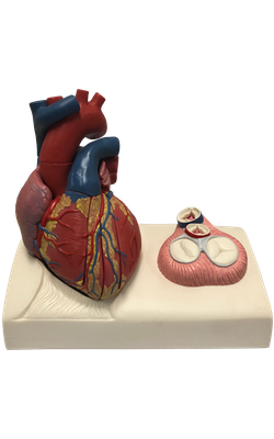
Main Model
Right atrium
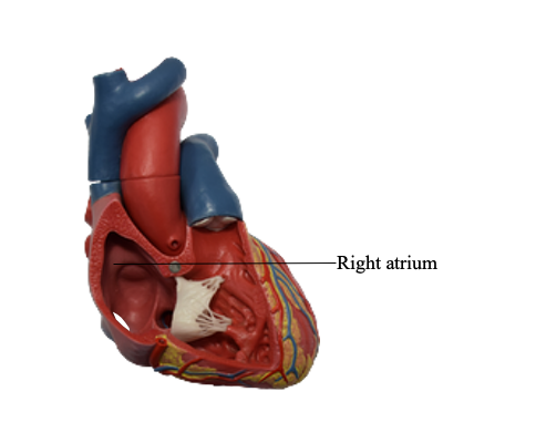
The right atrium forms the right border of the heart and receives venous blood from superior vena cava, inferior vena cava and coronary sinus. The ear-like right auricle is a conical muscular pouch that projects from this chamber like an add-on room, increasing the capacity of the atrium as it overlaps the ascending aorta.
The interior of the right atrium has a:
• Smooth, thin-walled, posterior part (the sinus venarum) on which the venae cavae (superior vena cava and inferior vena cava) and coronary sinus open, bringing poorly oxygenated blood into the heart.
• Rough, muscular anterior wall composed of pectinate muscles (Latin musculi pectinati).
• Right atrioventicular orifice through which the right atrium discharges the poorly oxygenated blood it has received into the right ventricle.
The smooth and rough parts of the atrial wall are separated externally by a shallow vertical groove, the sulcus terminalis or terminal groove, and internally by a vertical ridge, the crista terminalis or terminal crest. The superior vena cava opens into the superior part of the right atrium at the level of the right 3rd costal cartilage. The inferior vena cava opens into the inferior part of the right atrium almost in line with the superior vena cava at approximately the level of the 5th costal cartilage.