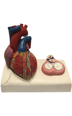
Main Model
Aortic valve (Left semilunar cusp)
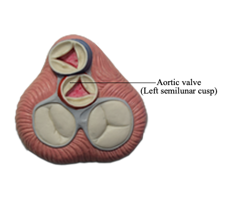
Semilunar Valve
Each of three semilunar cusps of the aortic valve (posterior, right and left) is concave when viewed superiorly. Semilunar cusps do not have tendinous cords to support them. They are smaller in area than the cusps of the atrioventricular valves, and the force exerted on them is less than half that exerted on the cusps of the tricuspid and mitral valves. The cusps project into the artery but are pressed toward (and not against) its walls as blood leaves the ventricle. After relaxation of the ventricle (diastole), the elastic recoil of the wall of the aorta forces the blood back toward the heart. However, the cusps snap closed like an umbrella caught in the wind as they catch the reversed blood flow. They come together to completely close the orifice, supporting each other as their edges abut (meet), and preventing any significant amount of blood from returning to the ventricle. The edge of each cusp is thickened in the region of contact, forming the lunule; the apex of the angulated free edge is thickened further as the nodule. Immediately superior to each semilunar cusp, the walls of the origins of the aorta are slightly dilated, forming a sinus. The aortic sinuses are the spaces at the origin of the ascending aorta between the dilated wall of the vessel and each cusp of the semilunar valves. The blood in the sinuses and the dilation of the wall prevent the cusps from sticking to the wall of the vessel, which might prevent closure.
The mouth of the right coronary artery is in the right aortic sinus, the mouth of the left coronary artery is in the left aortic sinus, and no artery arises from the posterior aortic (non-coronary) sinus.