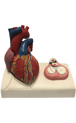
Main Model
Left ventricle
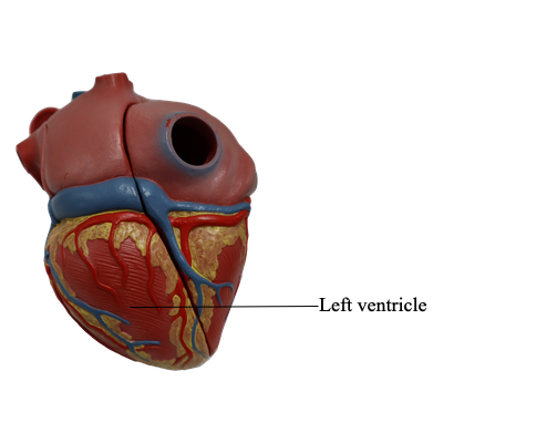
The left ventricle forms the apex of the heart, nearly all its left (pulmonary) surface and border, and most of the diaphragmatic surface. Because arterial pressure is much higher in the systemic than in the pulmonary circulation, the left ventricle performs more work than the right ventricle.
The interior of the left ventricle has
• Walls that are two to three times as thick as those of the right ventricle.
• Walls that are mostly covered with a mesh of trabeculae carneae that are finer and more numerous than those of the right ventricle.
• A conical cavity that is longer than that of the right ventricle.
• Anterior and posterior papillary muscles that are larger than those in the right ventricle.
• A smooth-walled, non-muscular, supero-anterior outflow part, the aortic vestibule, leading to the aortic orifice and aortic valve.
• A double-leaflet mitral valve that guards the left AV orifice.
• An aortic orifice that lies in its right posterosuperior part and is surrounded by a fibrous ring to which the right posterior, and left cusps of the aortic valve are attached; the ascending aorta begins at the aortic orifice.
The mitral valve has two cusps, anterior and posterior. The adjective mitral derives from the valve’s resemblance to a bishop’s miter (headdress). The mitral valve is located posterior to the sternum at the level of the 4th costal cartilage.
Each of its cusps receives tendinous cords from more than one papillary muscle. These muscles and their cords support the mitral valve, allowing the cusps to resist the pressure developed during contractions (pumping) of the left ventricle. The cords become taut just before and during systole, preventing the cusps from being forced into the left atrium. As it traverses the left ventricle, the bloodstream undergoes two right angle turns, which together result in a 180° change in direction. This reversal of flow takes place around the anterior cusp of the mitral valve.
The semilunar aortic valve, between the left ventricle and the ascending aorta, is obliquely placed. It is
located posterior to the left side of the sternum at the level of the 3rd intercostal space.