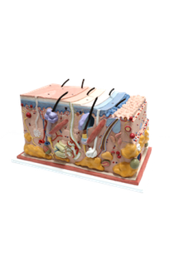
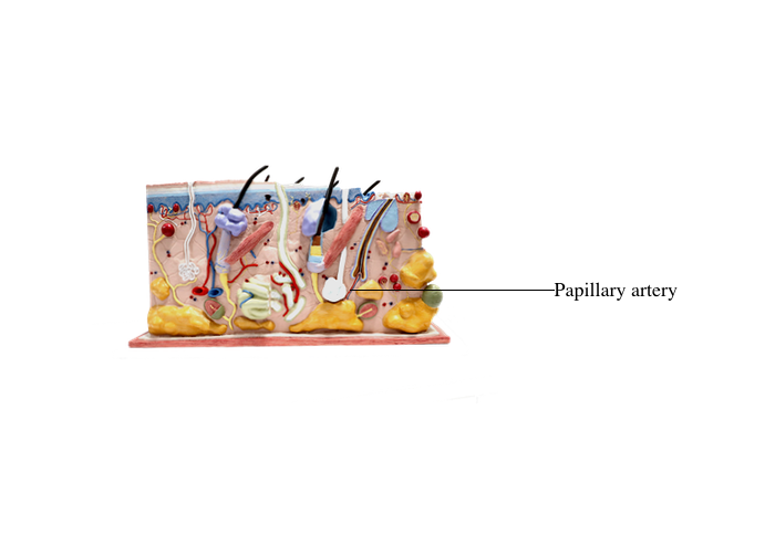
Main Model

Anterior : Papillary artery

Blood and Lymphatic Supply to the Skin
The cutaneous vascular supply has a primary function: thermoregulation. The secondary function is
nutrition of the skin and appendages. The arrangement of blood vessels permits rapid modification of
blood flow according to the required loss or conservation of heat.
Three interconnected networks are recognized in
the skin:
1. The subpapillary plexus, running along the
papillary layer of the dermis.
2. The cutaneous plexus, observed at the boundary
of the papillary and reticular layers of the dermis.
3. The hypodermic or subcutaneous plexus, present in the hypodermis or subcutaneous adipose tissue.
The subpapillary plexus gives rise to single loops of
capillaries within each dermal papilla. Venous blood
from the subpapillary plexus drains into veins of the
cutaneous plexus.
Branches of the hypodermic and cutaneous plexuses nourish the adipose tissue of the hypodermis,
the sweat glands, and the deeper segment of the hair
follicle.
Arteriovenous anastomoses (shunts) between the
arterial and venous circulation bypass the capillary
network. They are common in the reticular and
hypodermic regions of the extremities (hands, feet,
ears, lips, nose) and play a role in thermoregulation
of the body. The vascular shunts, under autonomic
vasomotor control, restrict flow through the superficial plexuses to reduce heat loss, ensuring deep cutaneous blood circulation. In some areas of the body
(for example, the face), cutaneous blood circulation
is also affected by an emotional state.
A special form of arteriovenous shunt is the glomus
apparatus. It is found in the dermis of the fingertips,
under the fingernails and toes and is involved in
temperature regulation. The glomus consists of an
endothelial-lined channel surrounded by cuboidal pericyte-like glomus cells and a rich nerve supply.
Glomus tumors are benign, usually very small (about
1 cm in diameter) red-blue nodules, associated with
sensitivity to cold and severe intermittent focal pain.
Surgical excision provides immediate pain relief.
Lymphatic vessels are blind endothelial cell-lined
spaces located below the papillary layer of the dermis,
collecting interstitial fluid for return to the blood
circulation. They also transport Langerhans cells to
regional lymph nodes.