Main Model
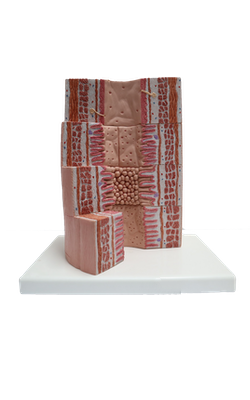
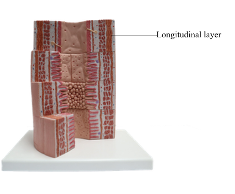
Esophagus [gullet] : Longitudinal layer
Esophagus
The esophagus is a muscular tube linking the pharynx to the stomach. It runs through the thorax, crosses the diaphragm, and enters the stomach. Contractions of the muscularis propel the food down the esophagus in about 2 seconds. At this velocity, changes of pressure and volume within the thorax are minimal. No disruption of respiration and cardiopulmonary circulation takes place.
The esophageal mucosa consists of a stratified squamous epithelium overlying a lamina propria with numerous connective tissue papillae. The muscularis mucosae is not present in the upper portion of the esophagus, but it becomes organized near the stomach.
The mucosa and the submucosa in the undistended esophagus form longitudinal folds that give the lumen an irregular outline. As the bolus of food moves down the esophagus, the folds disappear transiently and then are restored by the recoil of the elastic fibers of the submucosa.
The submucosa contains a network of collagen and elastic fibers and many small blood vessels. At the lower end of the esophagus, submucosal venous plexuses drain into both the systemic venous system and the portal venous system. An increase in pressure in the portal venous system, caused by chronic liver disease, results in dilation of the submucosal venous sinuses and the formation of esophageal varices. Rupture of the varices or ulceration of the overlying mucosa can produce hemorrhage into the esophagus and stomach, often causing vomiting (hematemesis).
Mucosal and submucosal glands are found in the esophagus. Their function is to produce continuously a thin layer of mucus that lubricates the surface of the epithelium.
The mucosal tubular glands, residing in the lamina propria, resemble the cardiac glands of the stomach and are called cardiac esophageal glands.
The submucosal tubuloacinar glands, found in the submucosa just beneath the muscularis mucosae, are organized into small lobules drained by a single duct. The acini are lined by two secretory cell types: a mucous and a serous cell type, the latter with secretory granules containing lysozyme.
The composition of the inner circumferential (or circular) and outer longitudinal layers of the muscularis shows segment-dependent variations. In the upper third of the esophagus, both layers consist of striated muscle. In the middle third, smooth muscle fibers can be seen deep to the striated muscle. In the lower third, both layers of the muscularis contain smooth muscle cells.
Clinical Significance: Barrett's Metaplasia
The esophagus has two sphincters:
1. The anatomically defined upper esophageal sphincter (UES), or cricopharyngeal sphincter.
2. The functionally defined lower esophageal sphincter (LES), or gastroesophageal sphincter.
The UES participates in the initiation of swallowing. The LES prevents reflux of gastric contents into the esophagus.
Because the esophageal stratified squamous lining epithelium at the epithelial transformation zone may be replaced at the lower end by a poorly resistant columnar epithelium (a process called Barrett's esophagus or metaplasia). Gastroesophageal reflux disease, (GERD) causes chronic inflammation or ulceration and difficulty in swallowing (dysphagia).
When the esophageal hiatus in the diaphragm does not close entirely during development, a hiatus hernia enables a portion of the stomach to move into the thoracic cavity. In sliding hiatus hernia, the stomach protrudes through the diaphragmatic hiatus, normally occupied by the lower esophagus.
GERD and peptic ulceration in the intrathoracic portion of the stomach and lower esophagus leads to difficulty in swallowing and the feeling of a lump in the throat. This condition, commonly seen in family practice patients, affects young and middle-aged women in particular.
The movements involved in swallowing are coordinated by nerves from the cervical and thoracic sympathetic trunks, forming plexuses in the submucosa and in between the inner and outer layers of the muscularis.
Diseases affecting this neuromuscular system may result in muscle spasm, difficulty in swallowing, and substernal pain.