Main Model
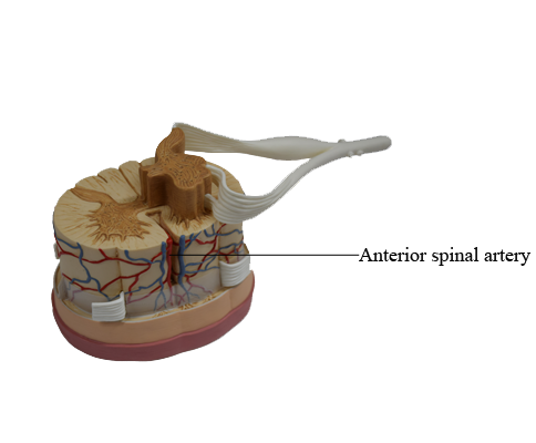
Anterior : Anterior spinal artery
Arteries of Spinal Cord and Nerve Roots
The arteries supplying the spinal cord are branches of the vertebral, ascending cervical, deep cervical, intercostal, lumbar, and lateral sacral arteries. Three longitudinal arteries supply the spinal cord: an anterior spinal artery and paired posterior spinal arteries. These arteries run longitudinally from the medulla of the brainstem to the conus medullaris of the spinal cord.
The anterior spinal artery, formed by the union of branches of the vertebral arteries, runs inferiorly in the anterior median fissure. Sulcal arteries arise from the anterior spinal artery and enter the spinal cord through this fissure. The sulcal arteries supply approximately two thirds of the cross-sectional area of the spinal cord.
Each posterior spinal artery is a branch of either the vertebral artery or the posteroinferior cerebellar artery. The posterior spinal arteries commonly form anastomosing channels in the pia mater.
By themselves, the anterior and posterior spinal arteries can supply only the short superior part of the spinal cord. The circulation to much of the spinal cord depends on segmental medullary and radicular arteries running along the spinal nerve roots. The anterior and posterior segmental medullary arteries are derived from spinal branches of the ascending cervical, deep cervical, vertebral, posterior intercostal, and lumbar arteries. The segmental medullary arteries occur mainly in association with the cervical and lumbosacral enlargements, regions where the need for a good blood supply is greatest. They enter the vertebral canal through the IV foramina.
The great anterior segmental medullary artery (of Adamkiewicz), which is on the left side in about 65% of people, reinforces the circulation to two thirds of the spinal cord, including the lumbosacral enlargement. The great artery, much larger than the other segmental medullary arteries, usually arises via a spinal branch from an inferior intercostal or upper lumbar artery and enters the vertebral canal through the IV foramen at the lower thoracic or upper lumbar level.
The posterior and anterior roots of the spinal nerves and their coverings are supplied by posterior and anterior radicular arteries (Latin radix, root), which run along the nerve roots. The radicular arteries do not reach the posterior, anterior, or spinal arteries. Segmental medullary arteries replace the radicular arteries at the irregular levels at which they occur. Most radicular arteries are small and supply only the nerve roots; however, some of them may assist with the supply of superficial parts of the gray matter in the posterior and anterior horns of the spinal cord.
Veins of Spinal Cord
In general, the veins of the spinal cord have a distribution similar to that of the spinal arteries. There are usually three anterior spinal veins and three posterior spinal veins. The spinal veins are arranged longitudinally, communicate freely with each other, and are drained by upto 12 anterior and posterior medullary and radicular veins. The veins of the spinal cord join the internal vertebral (epidural) venous plexuses in the epidural space. The internal vertebral venous plexuses pass superiorly through the foramen magnum to communicate with dural sinuses and vertebral veins in the cranium. The internal vertebral plexuses also communicate with the external vertebral venous plexuses on the external surface of the vertebrae.