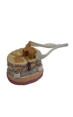
Main Model
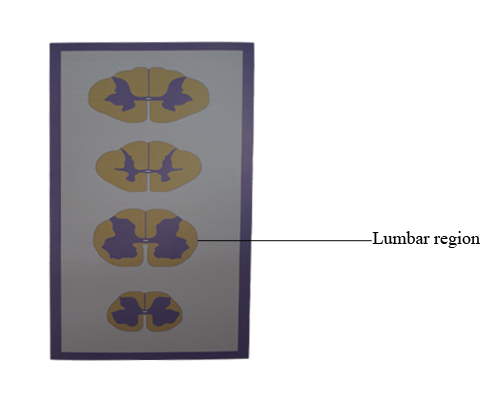
Top : Lumbar region
Regional Characteristics
Although all spinal levels have posterior, lateral, and anterior funiculi and posterior and anterior horns, their shapes and proportions vary between major spinal regions. For example, cervical (C4 to T1) and lumbosacral (L1 to S2) cord levels have prominent posterior and anterior horns because of the extensive sensory input from and motor outflow to the upper and lower extremities. In contrast, the posterior and anterior horns at thoracic levels are small, sensory input is less dense, and there is no appendicular musculature at these levels.
When viewing the spinal cord in the clinical setting, as in magnetic resonance imaging (MRI) or computed tomography (CT), it is important to note that anterior and posterior are reversed compared with the orientation commonly used in the anatomic setting. It is absolutely essential to remember the orientation and consequent location of tracts in the spinal cord in CT and MRI because these images are used in the evaluation of the neurologically compromised patient. In short, anterior is up in the image and posterior is down. In addition, the right and left sides of the patient are standardized; the observer's right is the patient's left and the observer's left is the patient's right.
Cervical Levels
The cervical cord is round to oval and proportionately larger than at other spinal levels. There is a large amount of white matter because a full complement of ascending and descending fiber tracts is present. The gracile and cuneate fasciculi are especially obvious structures at cervical levels.
At cervical levels C1 to C3, the posterior and anterior horns are comparatively small, which results in a more round than oval shape of the upper cervical spinal cord. The horns at C3 to C4 are becoming larger as part of the cervical enlargement; thus the shape of the spinal cord is transitioning from more round to more oval. At cervical levels C4 to C8, the posterior and anterior horns are large, reflecting the sensory input from and motor innervation to the upper extremity. At these levels, the spinal cord is distinctly oval. The oval shape of the spinal cord is also obvious in a myelogram at lower cervical levels.
Thoracic Levels
In general, the thoracic cord is round and the posterior and anterior horns are small. From upper to lower thoracic levels, there is a progressive decrease in the amount of white matter. Although both the gracile and cuneate fasciculi are present at upper thoracic levels (above T6), only the gracile fasciculus is present at lower thoracic levels (below T6). However, the small size of the posterior and anterior horns makes the white matter in thoracic levels appear proportionately large. As seen in a myelogram at about midthoracic levels, there is more space around the spinal cord than at lower cervical levels.
Two structures especially obvious in the gray matter of lamina VII at thoracic levels are the posterior thoracic nucleus (dorsal nucleus of Clarke) and the lateral horn. The posterior thoracic nucleus, a prominent medial cell group, contains neurons whose axons project to the cerebellum. The lateral horn is a protrusion into the lateral funiculus formed by the underlying intermediolateral cell column. These cells are preganglionic sympathetic neurons whose axons will terminate in either paravertebral or prevertebral ganglia.
Lumbar Levels
At lumbar levels the cord is also round. The posterior and anterior horns are large, and there is considerably less white matter than at higher levels. Therefore, the posterior and anterior horns appear proportionately large, the reverse of the situation at thoracic levels. In parallel with the situation at cervical levels, the proportionately large size of the posterior and anterior horns at lumbar levels accommodates, respectively, the significant sensory input from and motor outflow to the lower extremity. The posterior thoracic nucleus (dorsal nucleus of Clarke) is usually obvious at L1 and possibly L2 levels.
As the posterior and anterior roots descend in the more caudal portions of the dural sac, they form fascicles around the lower lumbar and sacral levels of the spinal cord. Myelograms at these levels clearly illustrate the position of the roots in relation to the spinal cord, and as they descend, they coalesce to form the cauda equina within the lumbar cistern.
Sacral Levels
At sacral levels, the spinal cord is round and smaller than at lumbar levels. It consists mainly of gray matter; the white matter forms a relatively thin shell. The intermediate gray matter at levels S2, S3, and S4 contains preganglionic parasympathetic cell bodies (the sacral visceromotor nucleus). The substantia gelatinosa (lamina II) is especially obvious at sacral levels.
At sacral levels the cord is small and surrounded by the posterior and anterior roots that are descending in the dural sac. At this point, the roots are passing caudal to form the cauda equina. The subarachnoid space around the lumbar, sacral, and coccygeal cord is continuous with the space of the lumbar cistern; a myelogram through the lumbar cistern reveals the roots forming the cauda equina but no spinal cord.