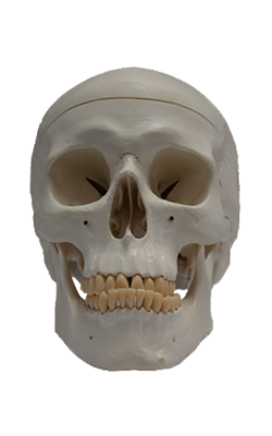
Main Model
CRANIUM : Right lateral aspect of cranium
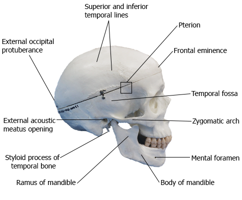
The temporal fossa is bounded superiorly and posteriorly by the superior and inferior temporal lines, anteriorly by the frontal and zygomatic bones, and inferiorly by the zygomatic arch. The superior border of this arch corresponds to the inferior limit of the cerebral hemisphere of the brain. The zygomatic arch is formed by the union of the temporal process of the zygomatic bone and the zygomatic process of the temporal bone.
In the anterior part of the temporal fossa, 3-4 cm superior
to the midpoint of the zygomatic arch, is a clinically important area of bone junctions: the pterion (Greek pteron, wing). It is usually indicated by an
H-shaped formation of sutures that unite the frontal, parietal, sphenoid (greater wing), and temporal bones. Less commonly, the frontal and temporal bones articulate; sometimes
all four bones meet at a point.
Landmark: Pterion (Greek wing)
Shape and Location: Junction of greater wing of sphenoid, squamous temporal, frontal, and parietal bones; overlies course of
anterior division of middle meningeal artery
The external acoustic meatus opening (pore) is the
entrance to the external acoustic meatus (canal), which leads to
the tympanic membrane (eardrum). The mastoid
process of the temporal bone is postero-inferior to the external acoustic meatus opening. Anteromedial to the mastoid process is the styloid process of the temporal bone, a slender
needle-like, pointed projection. The infratemporal fossa is an
irregular space inferior and deep to the zygomatic arch, and
the mandible and posterior to the maxilla.
The mandible is a U-shaped bone with an alveolar process that supports the mandibular teeth. It consists of a
horizontal part, the body, and a vertical part, the ramus. Inferior to the second premolar teeth are
the mental foramina for the mental nerves and vessels. The mental protuberance, forming the prominence of the chin, is a triangular
bony elevation inferior to the mandibular symphysis
(Latin symphysis menti), the osseous union where the halves of
the infantile mandible fuse.
The external occipital protuberance, is usually easily
palpable in the median plane; however, occasionally (especially in females) it may be inconspicuous. A craniometric
point defined by the tip of the external protuberance is the
inion (Greek nape of neck).