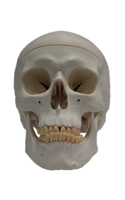
Main Model
CRANIUM : Cranial foramina
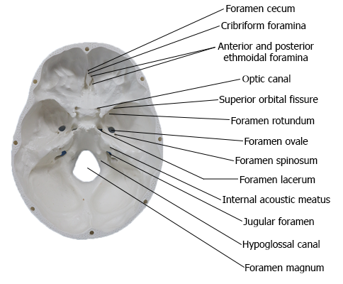
Internal Surface of Cranial Base
The internal surface of the cranial base (Latin basis cranii
interna) has three large depressions that lie at different levels: the anterior, middle, and posterior cranial fossae, which
form the bowl-shaped floor of the cranial cavity. The anterior cranial fossa is at the highest level, and the posterior cranial fossa is at the lowest level.
Anterior Cranial Fossa
The inferior and anterior parts of the frontal lobes of the brain
occupy the anterior cranial fossa, the shallowest of the three
cranial fossae. The fossa is formed by the frontal bone anteriorly, the ethmoid bone in the middle, and the body
and lesser wings of the sphenoid posteriorly. The greater part of
the fossa is formed by the orbital parts of the frontal bone, which support the frontal lobes of the brain and form the roofs of
the orbits. This surface shows sinuous impressions (brain markings) of the orbital gyri (ridges) of the frontal lobes.
The frontal crest is a median bony extension of the frontal bone. At its base is the foramen cecum of the frontal bone, which gives passage to vessels during fetal
development, but is insignificant postnatally. The crista galli
(Latin cock's comb) is a thick, median ridge of bone posterior
to the foramen cecum, which projects superiorly from the
ethmoid. On each side of this ridge is the sieve-like cribriform plate of ethmoid bone. Its numerous tiny foramina transmit the olfactory nerves (CN I) from the olfactory areas
of the nasal cavities to the olfactory bulbs of the brain, which
lie on this plate.
Middle Cranial Fossa
The butterfly-shaped middle cranial fossa has a central part
composed of the sella turcica on the body of the sphenoid and
large, depressed lateral parts on each side. The
middle cranial fossa is postero-inferior to the anterior cranial
fossa, separated from it by the sharp sphenoidal crests laterally
and the sphenoidal limbus centrally. The sphenoidal crests
are formed mostly by the sharp posterior borders of the lesser wings of the sphenoid bones, which overhang the lateral parts
of the fossae anteriorly. The sphenoidal crests end medially
in two sharp bony projections, the anterior clinoid processes.
A variably prominent ridge, the limbus of the sphenoid
forms the anterior boundary of the transversely oriented
prechiasmatic sulcus extending between the right and the
left optic canals. The bones forming the lateral parts of the
fossa are the greater wings of the sphenoid, and squamous
parts of the temporal bones laterally, and the petrous parts
of the temporal bones posteriorly. The lateral parts of the
middle cranial fossa support the temporal lobes of the brain.
The boundary between the middle and the posterior cranial
fossae is the superior border of the petrous part of the
temporal bone laterally, and a flat plate of bone, the dorsum sellae of the sphenoid, medially.
The sella turcica (Latin Turkish saddle) is the saddle-like
bony formation on the upper surface of the body of the sphenoid, which is surrounded by the anterior and posterior
clinoid processes. Clinoid means "bedpost," and the four processes (two anterior and two
posterior) surround the hypophysial fossa, the "bed" of the
pituitary gland, like the posts of a four-poster bed. The sella
turcica is composed of three parts:
1. The tuberculum sellae (horn of saddle): a variable slight
to prominent median elevation forming the posterior
boundary of the prechiasmatic sulcus and the anterior
boundary of the hypophysial fossa.
2. The hypophysial fossa (pituitary fossa): a median
depression (seat of saddle) in the body of the sphenoid
that accommodates the pituitary gland (Latin hypophysis).
3. The dorsum sellae (back of saddle): a square plate of
bone projecting superiorly from the body of the sphenoid.
It forms the posterior boundary of the sella turcica, and
its prominent superolateral angles make up the posterior
clinoid processes.
On each side of the body of the sphenoid, a crescent of four
foramina perforate the roots of the cerebral surfaces of the
greater wings of the sphenoids:
1. Superior orbital fissure: Located between the greater and
the lesser wings, it opens anteriorly into the orbit.
2. Foramen rotundum (round foramen): Located posterior
to the medial end of the superior orbital fissure, it runs a
horizontal course to an opening on the anterior aspect of
the root of the greater wing of the sphenoid into a bony formation between the sphenoid, the
maxilla, and the palatine bones, the pterygopalatine fossa.
3. Foramen ovale (oval foramen): A large foramen posterolateral to the foramen rotundum; it opens inferiorly into
the infratemporal fossa.
4. Foramen spinosum (spinous foramen): Located posterolateral to the foramen ovale and opens into the infratemporal
fossa in relationship to the spine of the sphenoid.
The foramen lacerum (lacerated or torn foramen) is not
part of the crescent of foramina. This ragged foramen lies posterolateral to the hypophysial fossa, and is an artifact of a
dried cranium. In life, it is closed by a cartilage
plate. Only some meningeal arterial branches and small veins
are transmitted vertically through the cartilage, completely
traversing this foramen. The internal carotid artery and its
accompanying sympathetic and venous plexuses pass across the superior aspect of the cartilage (i.e., pass over the foramen), and some nerves traverse it horizontally, passing to a
foramen in its anterior boundary.
Extending posteriorly and laterally from the foramen lacerum is a narrow groove for the greater petrosal nerve
on the anterosuperior surface of the petrous part of the temporal bone. There is also a small groove for the lesser petrosal
nerve.
Posterior Cranial Fossa
The posterior cranial fossa, the largest and deepest of
the three cranial fossae, lodges the cerebellum, pons, and
medulla oblongata. The posterior cranial fossa
is formed mostly by the occipital bone, but the dorsum sellae of the sphenoid marks its anterior boundary centrally, and the petrous and mastoid parts of the temporal bones contribute its anterolateral "walls."
From the dorsum sellae there is a marked incline, the
clivus, in the center of the anterior part of the fossa leading to the foramen magnum. Posterior to this large opening,
the posterior cranial fossa is partly divided by the internal
occipital crest into bilateral large concave impressions, the
cerebellar fossae. The internal occipital crest ends in the
internal occipital protuberance formed in relationship
to the confluence of the sinuses, a merging of dural venous sinuses.
Broad grooves show the horizontal course of the transverse sinus and the S-shaped sigmoid sinus. At the base of
the petrous ridge of the temporal bone is the jugular foramen, which transmits several cranial nerves in addition to the
sigmoid sinus that exits the cranium as the internal jugular
vein (IJV). Anterosuperior to the jugular foramen is the internal acoustic meatus for the facial (CN
VII) and vestibulocochlear nerves (CN VIII), and the labyrinthine artery. The hypoglossal canal for the hypoglossal
nerve (CN XII) is superior to the anterolateral margin of the
foramen magnum.