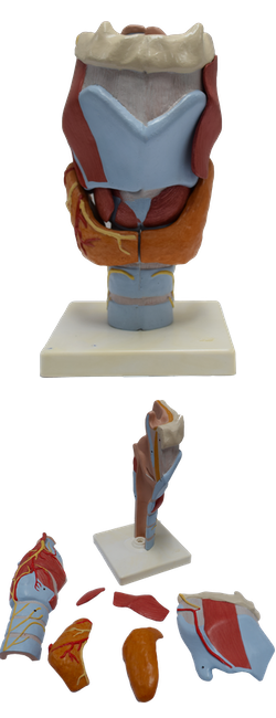
Main Model
Tracheal cartilages
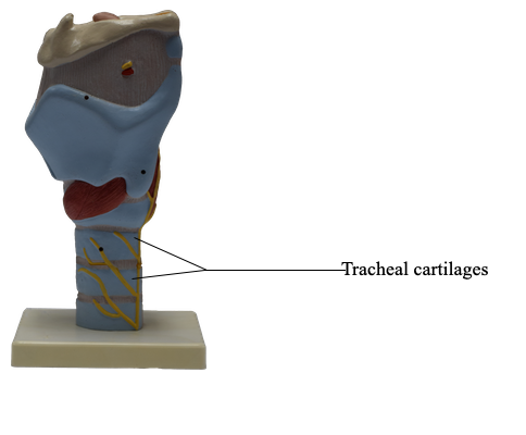
The trachea, extending from the larynx into the thorax, terminates inferiorly as it divides into right and left main bronchi. It transports air to and from the lungs, and its epithelium propels debris-laden mucus toward the pharynx for expulsion from the mouth. The trachea is a fibrocartilaginous tube, supported by incomplete cartilaginous tracheal cartilages (rings), that occupies a median position in the neck. The tracheal cartilages keep the trachea patent; they are deficient posteriorly where the trachea is adjacent to the esophagus. The posterior gaps in the tracheal rings are spanned by the involuntary trachealis muscle, smooth muscle connecting the ends of the rings. Hence the posterior wall of the trachea is flat.
In adults, the trachea is approximately 2.5 cm in diameter, whereas in infants it has the diameter of a pencil. The trachea extends from the inferior end of the larynx at the level of the C6 vertebra. It ends at the level of the sternal angle or the T4-T5 intervertebral disc, where it divides into the right and left main bronchi.
Lateral to the trachea are the common carotid arteries and the lobes of the thyroid gland. Inferior to the isthmus of the thyroid gland are the jugular venous arch and the inferior thyroid veins. The brachiocephalic trunk is related to the right side of the trachea in the root of the neck. Deviation of the trachea from the midline, apparent superficially or radiographically, often signals the presence of a pathological process. Tracheal trauma often affects the closely adherent esophagus.