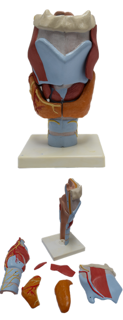
Main Model
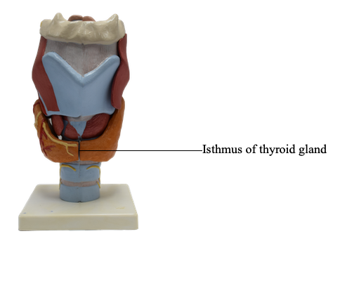
Isthmus of thyroid gland
The viscera of the endocrine layer are part of the body's endocrine system of ductless, hormone-secreting glands. The thyroid gland is the body's largest endocrine gland. It produces thyroid hormone, which controls the rate of metabolism, and calcitonin, a hormone controlling calcium metabolism. The thyroid gland affects all areas of the body except itself and the spleen, testes, and uterus. The hormone produced by the parathyroid glands, parathormone (PTH), controls the metabolism of phosphorus and calcium in the blood. The parathyroid glands target the skeleton, kidneys, and intestine.
The thyroid gland lies deep to the sternothyroid and sternohyoid muscles, located anteriorly in the neck at the level of the C5-T1 vertebrae. It consists primarily of right and left lobes, anterolateral to the larynx and trachea. A relatively thin isthmus unites the lobes over the trachea, usually anterior to the second and third tracheal rings. The thyroid gland is surrounded by a thin fibrous capsule, which sends septa deeply into the gland. Dense connective tissue attaches the capsule to the cricoid cartilage and superior tracheal rings. External to the capsule is a loose sheath formed by the visceral portion of the pretracheal layer of deep cervical fascia.
The highly vascular thyroid gland is supplied by the superior and inferior thyroid arteries. These vessels lie between the fibrous capsule and the loose fascial sheath. Usually the first branches of the external carotid arteries, the superior thyroid arteries, descend to the superior poles of the gland, pierce the pretracheal layer of deep cervical fascia, and divide into anterior and posterior branches supplying mainly the anterosuperior aspect of the gland.
The inferior thyroid arteries, the largest branches of the thyrocervical trunks arising from the subclavian arteries, run superomedially posterior to the carotid sheaths to reach the posterior aspect of the thyroid gland. They divide into several branches that pierce the pretracheal layer of the deep cervical fascia and supply the posteroinferior aspect, including the inferior poles of the gland. The right and left superior and inferior thyroid arteries anastomose extensively within the gland, ensuring its supply while providing potential collateral circulation between the subclavian and external carotid arteries.
In approximately 10% of people, a small, unpaired thyroid ima artery (Latin arteria thyroidea ima) arises from the brachiocephalic trunk; however, it may arise from the arch of the aorta or from the right common carotid, subclavian, or internal thoracic arteries. When present, this small artery ascends on the anterior surface of the trachea, supplying small branches to it. The artery then continues to the isthmus of the thyroid gland, where it divides and supplies it.
Three pairs of thyroid veins usually form a thyroid plexus of veins on the anterior surface of the thyroid gland and anterior to the trachea. The superior thyroid veins accompany the superior thyroid arteries; they drain the superior poles of the thyroid gland; the middle thyroid veins do not accompany but run essentially parallel courses with the inferior thyroid arteries; they drain the middle of the lobes. The usually independent inferior thyroid veins drain the inferior poles. The superior and middle thyroid veins drain into the IJVs; the inferior thyroid veins drain into the brachiocephalic veins posterior to the manubrium.
The lymphatic vessels of this gland run in the interlobular connective tissue, usually near the arteries; they communicate with a capsular network of lymphatic vessels. From here, the vessels pass initially to prelaryngeal, pretracheal, and paratracheal lymph nodes. The prelaryngeal nodes drain in turn to the superior deep cervical lymph nodes, and the pretracheal and paratracheal lymph nodes drain to the inferior deep cervical nodes. Laterally, lymphatic vessels located along the superior thyroid veins pass directly to the inferior deep cervical lymph nodes. Some lymphatic vessels may drain into the brachiocephalic lymph nodes or the thoracic duct.
The nerves of the thyroid gland are derived from the superior, middle, and inferior cervical (sympathetic) ganglia. They reach the gland through the cardiac and superior and inferior thyroid peri-arterial plexuses that accompany the thyroid arteries. These fibers are vasomotor, not secretomotor. They cause constriction of blood vessels. Endocrine secretion from the thyroid gland is hormonally regulated by the pituitary gland.