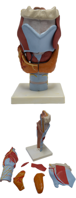
Main Model
Greater horn of hyoid bone
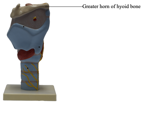
The mobile hyoid bone (or simply, the hyoid), lies in the anterior part of the neck at the level of the C3 vertebra in the angle between the mandible and the thyroid cartilage. The hyoid is suspended by muscles that connect it to the mandible, styloid processes, thyroid cartilage, manubrium of the sternum, and scapulae.
The hyoid is unique among bones for its isolation from the remainder of the skeleton. The U-shaped hyoid derives its name from the Greek word hyoeidçs, meaning “shaped like the letter upsilon,” the 20th letter in the Greek alphabet. The hyoid does not articulate with any other bone. It is suspended from the styloid processes of the temporal bones by the stylohyoid ligaments and is firmly bound to the thyroid cartilage. The hyoid consists of a body and greater and lesser horns (Latin cornua). Functionally, the hyoid serves as an attachment for anterior neck muscles and a prop to keep the airway open.
The body of the hyoid, its middle part, faces anteriorly and is approximately 2.5 cm wide and 1 cm thick. Its anterior convex surface projects anterosuperiorly; its posterior concave surface projects postero-inferiorly. Each end of its body is united to a greater horn that projects posterosuperiorly and laterally from the body. In young people, the greater horns are united to the body by fibrocartilage. In older people, the horns are usually united by bone. Each lesser horn is a small bony projection from the superior part of the body of the hyoid near its union with the greater horn. It is connected to the body of the hyoid by fibrous tissue and sometimes to the greater horn by a synovial joint. The lesser horn projects superoposteriorly toward the styloid process; it may be partly or completely cartilaginous in some adults.