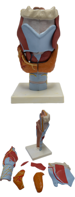
Main Model
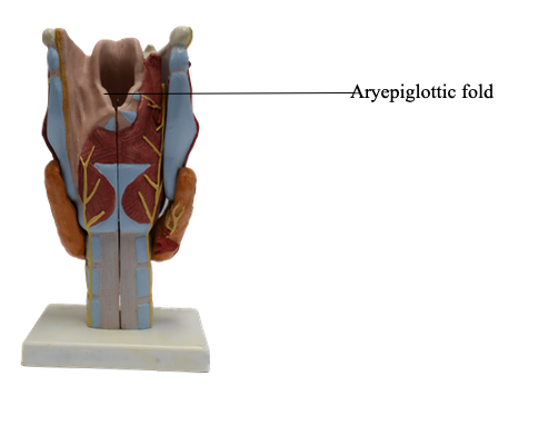
Aryepiglottic fold
The laryngeal skeleton consists of nine cartilages: three are single (thyroid, cricoid, and epiglottic), and three are paired (arytenoid, corniculate, and cuneiform).
The thyroid cartilage is the largest of the cartilages; its superior border lies opposite the C4 vertebra. The inferior two thirds of its two plate-like laminae fuse anteriorly in the median plane to form the laryngeal prominence. This projection (“Adam’s apple”) is well marked in men but seldom visible in women. Superior to this prominence, the laminae diverge to form a V-shaped superior thyroid notch. The less distinct inferior thyroid notch is a shallow indentation in the middle of the inferior border of the cartilage.
The posterior border of each lamina projects superiorly as the superior horn and inferiorly as the inferior horn. The superior border and superior horns attach to the hyoid by the thyrohyoid membrane. The thick median part of this membrane is the median thyrohyoid ligament; its lateral parts are the lateral thyrohyoid ligaments.
The inferior horns articulate with the lateral surfaces of the cricoid cartilage at the cricothyroid joints. The main movements at these joints are rotation and gliding of the thyroid cartilage, which result in changes in the length of the vocal folds. The cricoid cartilage is shaped like a signet ring with its band facing anteriorly. This ring-like opening of the cartilage fits an average finger. The posterior (signet) part of the cricoid is the lamina, and the anterior (band) part is the arch. Although much smaller than the thyroid cartilage, the cricoid cartilage is thicker and stronger and is the only complete ring of cartilage to encircle any part of the airway. It attaches to the inferior margin of the thyroid cartilage by the median cricothyroid ligament and to the first tracheal ring by the cricotracheal ligament. Where the larynx is closest to the skin and most accessible, the median cricothyroid ligament may be felt as a soft spot during palpation inferior to the thyroid cartilage.
The arytenoid cartilages are paired, three-sided pyramidal cartilages that articulate with the lateral parts of the superior border of the cricoid cartilage lamina. Each cartilage has an apex superiorly, a vocal process anteriorly, and a large muscular process that projects laterally from its base. The apex bears the corniculate cartilage and attaches to the ary-epiglottic fold. The vocal process provides the posterior attachment for the vocal ligament, and the muscular process serves as a lever to which the posterior and lateral crico-arytenoid muscles are attached. The crico-arytenoid joints, located between the bases of the arytenoid cartilages and the superolateral surfaces of the lamina of the cricoid cartilage, permit the arytenoid cartilages to slide toward or away from one to another, to tilt anteriorly and posteriorly, and to rotate. These movements are important in approximating, tensing, and relaxing the vocal folds.
The elastic vocal ligaments extend from the junction of the laminae of the thyroid cartilage anteriorly to the vocal process of the arytenoid cartilage posteriorly. The vocal ligaments make up the submucosal skeleton of the vocal folds. These ligaments are the thickened, free superior border of the conus elasticus or cricovocal membrane. The parts of the membrane extending laterally between the vocal folds and the superior border of the cricoid are the lateral cricothyroid ligaments. The fibroelastic conus elasticus blends anteriorly with the median cricothyroid ligament. The conus elasticus and overlying mucosa close the tracheal inlet except for the central rima glottidis (opening between the vocal folds).
The epiglottic cartilage, consisting of elastic cartilage, gives flexibility to the epiglottis, a heart-shaped cartilage covered with mucous membrane. Situated posterior to the root of the tongue and the hyoid and anterior to the laryngeal inlet, the epiglottic cartilage forms the superior part of the anterior wall and the superior margin of the inlet. Its broad superior end is free. Its tapered inferior end, the stalk of the epiglottis, is attached to the angle formed by the thyroid laminae by the thyro-epiglottic ligament. The hyo-epiglottic ligament attaches the anterior surface of the epiglottic cartilage to the hyoid. The quadrangular membrane is a thin, submucosal sheet of connective tissue that extends between the lateral aspects of the arytenoid and epiglottic cartilages. Its free inferior margin constitutes the vestibular ligament, which is covered loosely by mucosa to form the vestibular fold. This fold lies superior to the vocal fold and extends from the thyroid cartilage to the arytenoid cartilage. The free superior margin of the quadrangular membrane forms the ary-epiglottic ligament, which is covered with mucosa to form the ary-epiglottic fold. The corniculate and cuneiform cartilages appear as small nodules in the posterior part of the ary-epiglottic folds. The corniculate cartilages attach to the apices of the arytenoid cartilages; the cuneiform cartilages do not directly attach to other cartilages. The quadrangular membrane and conus elasticus are the superior and inferior parts of the submucosal fibroelastic membrane of the larynx.