Main Model
20 Feminine urethra

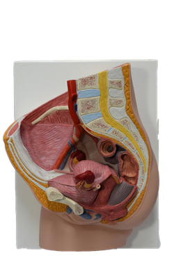
The female urethra (approximately 4 cm long and 6 mm in diameter) passes antero-inferiorly from the internal urethral orifice of the urinary bladder, posterior and then inferior to the pubic symphysis, to the external urethral orifice. The musculature surrounding the internal urethral orifice of the female bladder is not organized into an internal sphincter. The female external urethral orifice is located in the vestibule of the vagina, the cleft between the labia minora of the external genitalia, directly anterior to the vaginal orifice. The urethra lies anterior to the vagina (forming an elevation in the anterior vaginal wall); its axis is parallel to that of the vagina. The urethra passes with the vagina through the pelvic diaphragm, external urethral sphincter, and perineal membrane.
Urethral glands are present, particularly in the superior part of the urethra. One group of glands on each side, the paraurethral glands, are homologs to the prostate. These glands have a common para-urethral duct, which opens (one on each side) near the external urethral orifice. The external urethral sphincter is located in the perineum.
Blood is supplied to the female urethra by the internal pudendal and vaginal arteries. The veins follow the arteries and have similar names.
The nerves to the urethra arise from the vesical (nerve) plexus and the pudendal nerve. The pattern is similar to that in the male, given the absence of a prostatic plexus and an internal urethra sphincter. Visceral afferents from most of the urethra run in the pelvic splanchnic nerves, but the termination receives somatic afferents from the pudendal nerve. Both the visceral and the somatic afferent fibers extend from cell bodies in the S2-S4 spinal ganglia.