Main Model
50 Uterus

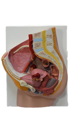
The uterus (womb) is a thick-walled, pear-shaped, hollow muscular organ. The embryo and fetus develop in the uterus. Its muscular walls adapt to the growth of the fetus and then provide the power for its expulsion during childbirth. The non-gravid (non-pregnant) uterus usually lies in the lesser pelvis, with its body lying on the urinary bladder and its cervix between the urinary bladder and rectum.
The adult uterus is usually anteverted (tipped anterosuperiorly relative to the axis of the vagina) and anteflexed (flexed or bent anteriorly relative to the cervix, creating the angle of flexion) so that its mass lies over the bladder. Consequently, when the bladder is empty, the uterus typically lies in a nearly transverse plane. The position of the uterus changes with the degree of fullness of the bladder and rectum, and stage of pregnancy. Although its size varies considerably, the non-gravid uterus is approximately 7.5 cm long, 5 cm wide, and 2 cm thick and weighs approximately 90 grams.
The uterus is divisible into two main parts: the body and cervix.
The body of the uterus, forming the superior two thirds of the organ, includes the fundus of the uterus, the rounded part that lies superior to the uterine ostia. The body lies between the layers of the broad ligament and is freely movable. It has two surfaces: vesical (related to the bladder) and intestinal. The body is demarcated from the cervix by the isthmus of the uterus, a relatively constricted segment, approximately 1 cm long.
The cervix of the uterus is the cylindrical, relatively narrow inferior third of the uterus, approximately 2.5 cm long in an adult non-pregnant woman. For descriptive purposes, two parts are described: a supravaginal part between the isthmus and the vagina, and a vaginal part, which protrudes into the superiormost anterior vaginal wall. The rounded vaginal part surrounds the external os of the uterus and is surrounded in turn by a narrow recess, the vaginal fornix. The supravaginal part is separated from the bladder anteriorly by loose connective tissue and from the rectum posteriorly by the recto-uterine pouch.
The slit-like uterine cavity is approximately 6 cm in length from the external os to the wall of the fundus. The uterine horns (Latin cornua) are the superolateral regions of the uterine cavity, where the uterine tubes enter. The uterine cavity continues inferiorly as the cervical canal. The fusiform canal extends from a narrowing inside the isthmus of the uterine body, the anatomical internal os, through the supravaginal and vaginal parts of the cervix, communicating with the lumen of the vagina through the external os. The uterine cavity (in particular, the cervical canal) and the lumen of the vagina together constitute the birth canal, through which the fetus passes at the end of gestation.
The wall of the body of the uterus consists of three coats, or layers:
• Perimetrium - the serosa or outer serous coat - consists of peritoneum supported by a thin layer of connective tissue.
• Myometrium - the middle coat of smooth muscle - becomes greatly distended (more extensive but much thinner) during pregnancy. The main branches of the blood vessels and nerves of the uterus are located in this coat. During childbirth, contraction of the myometrium is hormonally stimulated at intervals of decreasing length to dilate the cervical os and expel the fetus and placenta. During the menses, myometrial contractions may produce cramping.
• Endometrium - the inner mucous coat - is firmly adhered to the underlying myometrium. The endometrium is actively involved in the menstrual cycle, differing in structure with each stage of the cycle. If conception occurs, the blastocyst becomes implanted in this layer; if conception does not occur, the inner surface of this coat is shed during menstruation.
The amount of muscular tissue in the cervix is markedly less than in the body of the uterus. The cervix is mostly fibrous and is composed mainly of collagen with a small amount of smooth muscle and elastin.
The blood supply of the uterus derives mainly from the uterine arteries, with potential collateral supply from the ovarian arteries. The uterine veins enter the broad ligaments with the arteries and form a uterine venous plexus on each side of the cervix. Veins from the uterine plexus drain into the internal iliac veins.