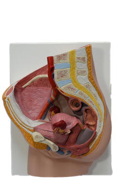
Main Model
35 Left ureter
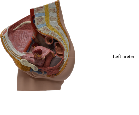
The ureters are muscular tubes, 25-30 cm long, that connect the kidneys to the urinary bladder. The ureters are retroperitoneal. As the ureters cross the bifurcation of the common iliac artery (or the beginning of the external iliac artery), they pass over the pelvic brim, thus leaving the abdomen and entering the lesser pelvis. The pelvic parts of the ureters run on the lateral walls of the pelvis, parallel to the anterior margin of the greater sciatic notch, between the parietal pelvic peritoneum and the internal iliac arteries. Opposite the ischial spine, they curve anteromedially, superior to the levator ani, and enter the urinary bladder. The inferior ends of the ureters are surrounded by the vesical venous plexus.
The ureters pass obliquely through the muscular wall of the urinary bladder in an inferomedial direction, entering the outer surface of the bladder approximately 5 cm apart, but their internal openings into the lumen of the empty bladder are separated by only half that distance. This oblique passage through the bladder wall forms a one-way “flap valve,” the internal pressure of the filling bladder causing the intramural passage to collapse. In addition, contractions of the bladder musculature act as a sphincter preventing the reflux of urine into the ureters when the bladder contracts, increasing internal pressure during micturition. Urine is transported down the ureters by means of peristaltic contractions, a few drops being transported at intervals of 12-20 seconds.
In males, the only structure that passes between the ureter and the peritoneum is the ductus deferens; it crosses the ureter within the ureteric fold of peritoneum. The ureter lies posterolateral to the ductus deferens and enters the posterosuperior angle of the bladder, just superior to the seminal gland.
In females, the ureter passes medial to the origin of the uterine artery and continues to the level of the ischial spine, where it is crossed superiorly by the uterine artery. It then passes close to the lateral part of the fornix of the vagina and enters the posterosuperior angle of the bladder.
The arterial supply to the pelvic parts of the ureters is variable, with ureteric branches extending from the common iliac, internal iliac, and ovarian arteries. The ureteric branches anastomose along the length of the ureter forming a continuous blood supply, although not necessarily effective collateral pathways. The most constant arteries supplying the terminal parts of the ureter in females are branches of the uterine arteries. The source of similar branches in males are the inferior vesical arteries. The blood supply of the ureters is a matter of great concern to surgeons operating in the region.
The venous drainage from the pelvic parts of the ureters generally parallels the arterial supply, draining to veins with corresponding names.
Lymphatic vessels pass primarily to common and internal iliac nodes.
The nerves to the ureters derive from adjacent autonomic plexuses (renal, aortic, superior and inferior hypogastric). The ureters are primarily superior to the pelvic pain line. Afferent (pain) fibers from the ureters follow sympathetic fibers in a retrograde direction to reach the spinal ganglia and spinal cord segments of T10-L2 or L3. Ureteric pain is usually referred to the ipsilateral lower quadrant of the abdomen, especially to the groin (inguinal region).