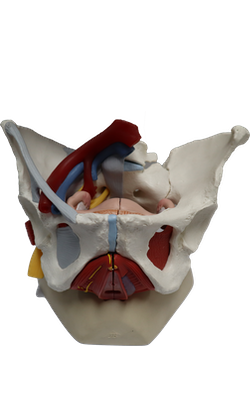
Main Model
ARTERIES : Internal iliac artery
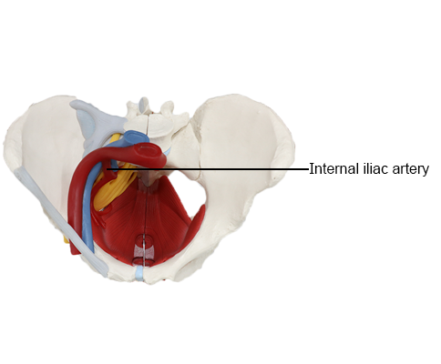
Internal Iliac Artery
The internal iliac artery is the principal artery of the pelvis,
supplying most of the blood to the pelvic viscera and some to
the musculoskeletal part of the pelvis; however, it also supplies branches to the gluteal region, medial thigh regions,
and the perineum.
Each internal iliac artery, approximately 4 cm long, begins
as the common iliac artery and bifurcates into the internal
and external iliac arteries at the level of the IV disc between
the L5 and S1 vertebrae. The ureter crosses the common
iliac artery or its terminal branches at or immediately distal
to the bifurcation. The internal iliac artery is separated from
the sacro-iliac joint by the internal iliac vein and the lumbosacral trunk. It descends posteromedially into the lesser
pelvis, medial to the external iliac vein and obturator nerve,
and lateral to the peritoneum.
Anterior Division of Internal Iliac Artery
Although
variations are common, the internal iliac artery usually
ends at the superior edge of the greater sciatic foramen by
dividing into anterior and posterior divisions (trunks). The branches of the anterior division of the internal iliac
artery are mainly visceral (i.e., they supply the bladder,
rectum, and reproductive organs), but they also include
parietal branches that pass to the thigh and buttocks. The arrangement of the visceral branches
is variable.
Umbilical Artery
Before birth, the umbilical arteries
are the main continuation of the internal iliac arteries, passing along the lateral pelvic wall and then ascending the anterior abdominal wall to and through the umbilical ring into
the umbilical cord.
Prenatally, the umbilical arteries conduct oxygen- and
nutrient-deficient blood to the placenta for replenishment.
When the umbilical cord is cut, the distal parts of these
vessels no longer function and become occluded distal
to branches that pass to the bladder. The occluded parts form fibrous cords called the medial umbilical ligaments. The ligaments raise folds of peritoneum (medial umbilical folds) on the deep surface of the
anterior abdominal wall.
Postnatally, the patent parts of the umbilical arteries run
antero-inferiorly between the urinary bladder and the lateral
wall of the pelvis.
Obturator Artery
The origin of the obturator artery
is variable; usually it arises close to the origin of the umbilical artery, where it is crossed by the ureter. It runs antero-inferiorly on the obturator fascia on the lateral wall of the
pelvis, and passes between the obturator nerve and vein.
Within the pelvis, the obturator artery gives off muscular
branches, a nutrient artery to the ilium, and a pubic branch.
The pubic branch arises just before the obturator artery
leaves the pelvis. It ascends on the pelvic surface of the pubis
to anastomose with its fellow of the opposite side, and the
pubic branch of the inferior epigastric artery, a branch of the
external iliac artery.
In a common variation (20%), an aberrant or accessory
obturator artery arises from the inferior epigastric artery
and descends into the pelvis along the usual route of the
pubic branch. Surgeons performing
hernia repairs must keep this common variation in mind.
Inferior Vesical Artery
The inferior vesical artery
occurs only in males, being replaced
by the vaginal artery in females.
Uterine Artery
The uterine artery is an additional
branch of the internal iliac artery in females, usually arising separately and directly from the internal iliac artery. It may arise from the umbilical artery. Developmentally, it is the homolog of the artery to the ductus deferens
in the male. It descends on the lateral wall of the pelvis, anterior to the internal iliac artery, and passes medially to reach the
junction of the uterus and vagina, where the cervix (neck) of
the uterus protrudes into the superior vagina.
As it passes medially, the uterine artery passes directly superior to the ureter. The relationship of ureter to artery is often
remembered by the phrase "water (urine) passes under the
bridge (uterine artery)." On reaching the side of the cervix,
the uterine artery divides into a smaller descending vaginal
branch, which supplies the cervix and vagina, and a larger
ascending branch, which runs along the lateral margin of
the uterus, supplying it. The ascending branch bifurcates into ovarian and tubal branches, which continue to supply the
medial ends of the ovary and uterine tube, and anastomose
with the ovarian and tubal branches of the ovarian artery.
Vaginal Artery
The vaginal artery is the homolog to
the inferior vesical artery in males. It often arises from the initial part of the uterine artery instead of arising directly from
the anterior division. The vaginal artery supplies numerous
branches to the anterior and posterior surfaces of the vagina.
Middle Rectal Artery
The middle rectal artery may
arise independently from the internal iliac artery, or it may
arise in common with the inferior vesical artery or the internal pudendal artery.
Internal Pudendal Artery
The internal pudendal
artery, larger in males than in females, passes inferolaterally, anterior to the piriformis muscle and sacral plexus. It leaves the pelvis between the piriformis and coccygeus muscles by passing through the inferior part of the greater sciatic
foramen. The internal pudendal artery then passes around
the posterior aspect of the ischial spine or the sacrospinous
ligament and enters the ischio-anal fossa through the lesser
sciatic foramen.
The internal pudendal artery, along with the internal
pudendal veins and branches of the pudendal nerve, passes
through the pudendal canal in the lateral wall of the ischio-anal fossa. As it exits the pudendal canal, medial
to the ischial tuberosity, the internal pudendal artery divides
into its terminal branches, the perineal artery and dorsal
arteries of the penis or clitoris.
Inferior Gluteal Artery
The inferior gluteal artery is
the larger terminal branch of the anterior division of the internal iliac artery, but may arise from the posterior division. It passes posteriorly between the sacral nerves (usually S2 and S3), and leaves the pelvis through the
inferior part of the greater sciatic foramen, inferior to the
piriformis muscle. It supplies the muscles and skin
of the buttock, and the posterior surface of the thigh.
Posterior Division of Internal Iliac Artery
When the
internal iliac artery divides into anterior and posterior divisions, the posterior division typically gives rise to the following three parietal arteries:
• Iliolumbar artery: This artery runs superolaterally in a
recurrent fashion (turning sharply backward relative to
its source) to the iliac fossa. Within the fossa, the artery
divides into an iliac branch, which supplies the iliacus
muscle and ilium, and a lumbar branch, which supplies the psoas major and quadratus lumborum muscles.
• Lateral sacral arteries: Superior and inferior lateral
sacral arteries may arise as independent branches or via
a common trunk. The lateral sacral arteries pass medially
and descend anterior to the sacral anterior rami, giving off
spinal branches, which pass through the anterior sacral foramina, and supply the spinal meninges enclosing the
roots of the sacral nerves. Some branches of these arteries pass from the sacral canal through the posterior sacral foramina and supply the erector spinae muscles of the
back and the skin overlying the sacrum.
• Superior gluteal artery: The largest branch of the posterior division, the superior gluteal artery supplies the gluteal muscles in the buttocks.