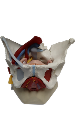
Main Model
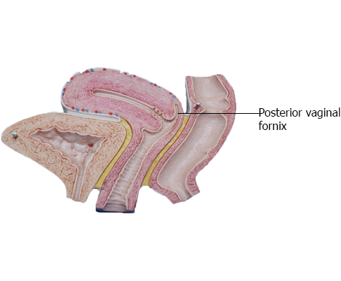
FEMALE INTERNAL GENITAL ORGANS : Posterior vaginal fornix
Vagina
The vagina, a distensible musculomembranous tube (7-9 cm long), extends from the middle cervix of the uterus to the vaginal orifice, the opening at the inferior end of the vagina. The vaginal orifice, external urethral orifice, and ducts of the greater and lesser vestibular glands open into the vestibule of the vagina, the cleft between the labia minora. The vaginal part of the cervix lies anteriorly in the superior vagina. The vagina:
• serves as a canal for menstrual fluid,
• forms the inferior part of the birth canal,
• receives the penis and ejaculate during sexual intercourse, and
• communicates superiorly with the cervical canal and inferiorly with the vestibule of the vagina.
The vagina is usually collapsed. The orifice is usually collapsed toward the midline so that its lateral walls are in contact on each side of an anteroposterior slit. Superior to the orifice, however, the anterior and posterior walls are in contact on each side of a transverse potential cavity, H-shaped in cross section, except at its superior end where the cervix holds them apart. The vagina lies posterior to the urinary bladder and urethra, the latter projecting along the midline of its inferior anterior wall. The vagina lies anterior to the rectum, passing between the medial margins of the levator ani (puborectalis) muscles. The vaginal fornix, the recess around the cervix, has anterior, posterior, and lateral parts. The posterior vaginal fornix is the deepest part and is closely related to the recto-uterine pouch. Four muscles compress the vagina and act as sphincters: pubovaginalis, external urethral sphincter, urethrovaginal sphincter, and bulbospongiosus.
The vagina is related:
• anteriorly to the fundus of the urinary bladder and urethra;
• laterally to the levator ani, visceral pelvic fascia, and ureters; and
• posteriorly (from inferior to superior) to the anal canal, rectum, and recto-uterine pouch.
Arterial Supply and Venous Drainage of Vagina
The arteries supplying the superior part of the vagina derive from the uterine arteries.The arteries supplying the middle and inferior parts of the vagina derive from the vaginal and internal pudendal arteries.
The vaginal veins form vaginal venous plexuses along the sides of the vagina and within the vaginal mucosa. These veins are continuous with the uterine venous plexus as the uterovaginal venous plexus, and drain into the internal iliac veins through the uterine vein. This plexus also communicates with the vesical and rectal venous plexuses.