Main Model
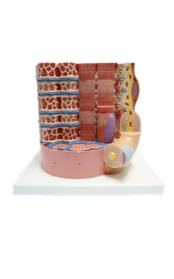
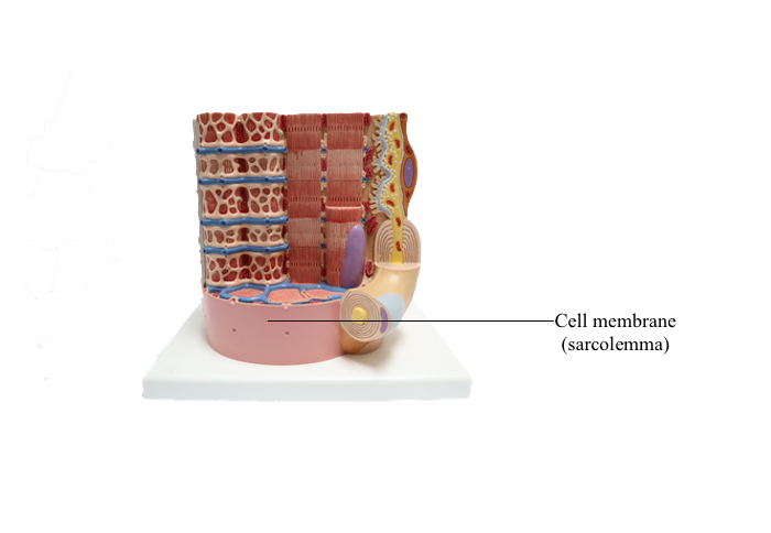
Anterior : Cell membrane (sarcolemma)
Characteristics of the Skeletal Muscle Cell or Fiber
Skeletal muscle cells are formed in the embryo by the fusion of myoblasts that produce a postmitotic, multinucleated myotube. The myotube matures into the long muscle cell with a diameter of 10 to 100 micrometer and a length of up to several centimeters.
The plasma membrane of the muscle cell (called the sarcolemma) is surrounded by a basal lamina and satellite cells.
The sarcolemma projects long, finger-like processes, called transverse tubules or T tubules, into the cytoplasm of the cell, the sarcoplasm. T tubules make contact with membranous sacs or channels, the sarcoplasmic reticulum.
The sarcoplasmic reticulum contains high concentrations of Ca2+. The site of contact of the T tubule with the sarcoplasmic reticulum cisternae is called a triad because it consists of two lateral sacs of the sarcoplasmic reticulum and a central T tubule.
The many nuclei of the muscle fiber are located at the periphery of the cell, just under the sarcolemma.
About 80% of the sarcoplasm is occupied by myofibrils surrounded by mitochondria (called sarcosomes). Myofibrils are composed of two major filaments formed by contractile proteins: thin filaments contain actin, and thick filaments are composed of myosin.
Depending on the type of muscle, mitochondria may be found parallel to the long axis of the myofibrils, or they may wrap around the zone of thick filaments. Thin filaments insert into each side of the Z disk (also called band, or line) and extend from the Z disk into the A band, where they alternate with thick filaments.