Main Model
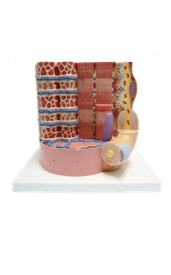
Anterior : Myofibrils
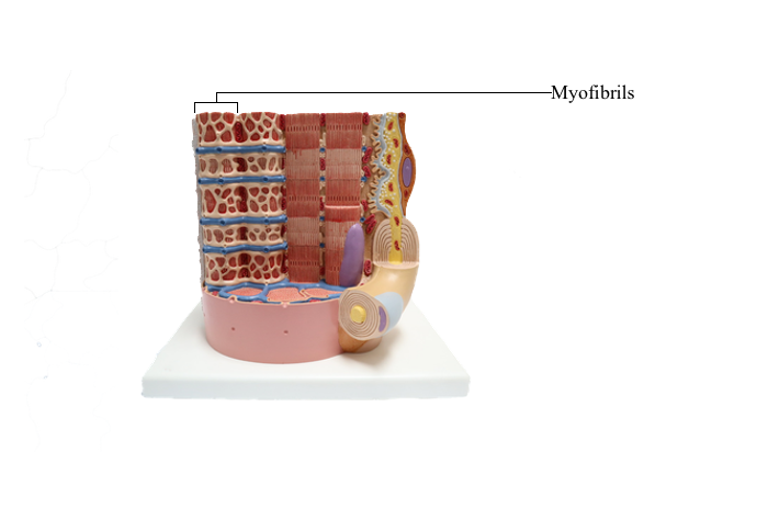
Muscular Tissue
Muscular tissue is specialized for contractility. It is composed of elongated, contractile muscle cells, which are often referred to as muscle fibers. Each muscle fiber contains numerous elongated, threadlike structures, myofibrils, which are composed of contractile proteins and are the smallest units of contraction. In muscular tissue, the muscle fibers usually are arranged in bundles, with their long axes parallel to one another. The muscular tissue is provided with a network of capillaries and nerve endings, which conduct the impulse from or toward the central nervous system.
There are three types of muscular tissue distinguished on both a structural and functional basis: smooth muscle, skeletal muscle, and cardiac muscle.
1. Smooth muscle is composed of bundles of fusiform muscle cells that do not show any striations under the light microscope. It forms the muscular component in the walls of viscera. The smooth muscle fibers may also function as single contractile units, such as the myoepithelial cells surrounding the acini of exocrine glands. Their contraction is slow and involuntary.
2. Skeletal muscle consists of bundles of elongated, cylindrical multinucleated muscle cells that show prominent cross striations. The skeletal muscle, attached to bones or fascia, is responsible for the movement of the skeleton and organs such as the tongue and eyeball. Their contraction is controlled by the will, and therefore is quick, forceful, and voluntary.
3. Cardiac muscle is formed of cylindrical and branching muscle cells, which also show cross striations. Cardiac muscle occurs in the wall of the heart and extends into the major veins opening into the heart. The contraction of the cardiac muscle is rhythmic, automatic, and involuntary.
Smooth Muscle
Smooth muscle is composed of long spindle-shaped cells, the smooth muscle fibers. These fibers are enveloped in fine fibroelastic tissue, forming the contractile portion of many organs. A single smooth muscle fiber is 20-500 micrometer long and 5-10 micrometer wide, with an elongated ovoid nucleus in the middle of the cell. In longitudinal section, the smooth muscle fibers are seen arranged with the nuclear (widest) region of one cell aligned with the tapering ends of its adjacent fibers. The cell processes of smooth muscle fibers can be clearly recognized in good specimens. These processes are short, narrow fingerlike projections, which make contact with adjacent smooth muscle cells. In cross section, the nucleus is round and is seen only in a few cell profiles.
Smooth muscle, also called visceral muscle, occurs in the digestive tract, blood vessels, lymphatic vessels, respiratory system, urinary system, reproductive system, dermis, and iris and ciliary body of the eye. Smooth muscle is innervated by autonomic (sympathetic and parasympathetic) nerve fibers, and exhibits a slow and continuing contraction. In the digestive tract, ganglion cells surrounded by satellite cells, a cell type of neuroglia in the peripheral nervous system, occur between two layers of smooth muscle.
Smooth muscle enables the viscera to contract in a rhythmic or wavelike fashion for long periods without fatigue. For example, the continuous rhythmic contractions of the smooth muscle in the gastrointestinal tract propel the luminal contents distantly.
Skeletal Muscle
Skeletal muscle consists of bundles of long cylindrical multinucleated muscle fibers (cells) with cross striations. The cells are 1-40 mm long and 10-100 micrometer wide. A single fiber may contain several hundred elongated ovoid nuclei at the periphery of the cell. At high magnification under the light microscope, the striations can be seen composed of alternate light and dark bands. The dark bands are termed A bands (anisotropic); the light bands are termed I bands (isotropic). In the middle of each I band is a dark line called the Z band or Z line parallel to the A and I bands. The distance between two Z lines is called a sarcomere, which is the structural and functional unit of the muscle contractile apparatus. Individual muscle fibers are covered by a thin layer of delicate connective tissue called endomysium, composed of a mixture of collagen fibers, elastic fibers, and fibroblasts. Between muscle fibers there is an abundance of capillaries and some nerve fibers.
Skeletal muscle, also called voluntary muscle, is innervated by large motor nerves through neuromuscular junctions, the motor end plate. The contraction of skeletal muscle is controlled voluntarily and is abrupt and strong yet transient (fatiguable).
Cardiac Muscle
Cardiac muscle is composed of striated muscle cells that contract involuntarily, rhythmically, and automatically. It is found only in the muscle layer of the heart (myocardium) and in the major veins joining the heart.
The muscle fibers are cylindrical, having an average length of 100-150 micrometer and width of 15-20 micrometer. A single muscle fiber has one or two elongated or ovoid nuclei, located centrally. Around the nuclei are fusiform areas of sarcoplasm containing many organelles. The cross striations formed with the contractile apparatus are less distinct than those in skeletal muscle fibers. Cardiac muscle fibers branch, bifurcate, and anastomose to form a complex three-dimensional network constituting the myocardium. Muscle fibers including their branches join end-to-end, with distinctive junctional zones marked by step like intercalated discs. Between muscle fibers is a layer of connective tissue containing collagen fibers, reticular fibers, and capillaries as well as fibroblasts.
Purkinje fibers are just beneath the subendothelial layer of the endocardium. These are specialized cardiac muscle fibers belonging to the impulse-conducting system of the heart. As cardiac muscle fibers, Purkinje fibers contain cross striations and are marked with intercalated discs at the interface of two fibers. However, they are larger and thicker (about 50 micrometer in diameter) than the usual cardiac muscle fibers and have very few myofibrils and much more central sarcoplasm.