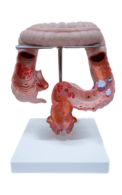
Main Model
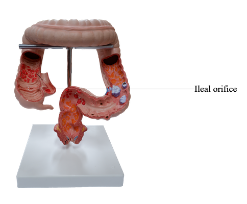
Ileal orifice
Cecum and Appendix
The cecum is the first part of the large intestine; it is continuous with the ascending colon. The cecum is a blind intestinal pouch, approximately 7.5 cm in both length and breadth. It lies in the iliac fossa of the right lower quadrant of the abdomen, inferior to the junction of the terminal ileum and cecum. If distended with feces or gas, the cecum may be palpable through the anterolateral abdominal wall.
The cecum usually lies within 2.5 cm of the inguinal ligament; it is almost entirely enveloped by peritoneum and can be lifted freely. However, the cecum has no mesentery. Because of its relative freedom, it may be displaced from the iliac fossa, but it is commonly bound to the lateral abdominal wall by one or more cecal folds of peritoneum. The terminal ileum enters the cecum obliquely and partly invaginates into it.
In dissection, the ileal orifice enters the cecum between ileocolic lips (superior and inferior), folds that meet laterally forming ridges called the frenula of the ileal orifice. It was believed that when the cecum is distended or when it contracts, the lips and frenula actively tighten, closing the valve to prevent reflux from the cecum into the ileum. However, direct observation by endoscopy in living persons does not support this description. The circular muscle is poorly developed around the orifice; therefore, the valve is unlikely to have any sphincteric action that controls passage of the intestinal contents from the ileum into the cecum. The orifice is usually closed by tonic contraction, however, appearing as an ileal papilla on the cecal side. The papilla probably serves as a relatively passive flap valve, preventing reflux from the cecum into the ileum as contractions occur to propel contents up the ascending colon and into the transverse colon.
The appendix (vermiform appendix; Latin vermis, worm-like) is a blind intestinal diverticulum (6-10 cm in length) that contains masses of lymphoid tissue. It arises from the posteromedial aspect of the cecum inferior to the ileocecal junction. The appendix has a short triangular mesentery, the meso-appendix, which derives from the posterior side of the mesentery of the terminal ileum. The meso-appendix attaches to the cecum and the proximal part of the appendix. The position of the appendix is variable, but it is usually retrocecal.
The arterial supply of the cecum is from the ileocolic artery, the terminal branch of the SMA. The appendicular artery, a branch of the ileocolic artery, supplies the appendix. Venous drainage from the cecum and appendix flow through a tributary of the SMV, the ileocolic vein.
Lymphatic drainage of the cecum and appendix passes to lymph nodes in the meso-appendix and to the ileocolic lymph nodes that lie along the ileocolic artery. Efferent lymphatic vessels pass to the superior mesenteric lymph nodes.
The nerve supply to the cecum and appendix derives from the sympathetic and parasympathetic nerves from the superior mesenteric plexus. The sympathetic nerve fibers originate in the lower thoracic part of the spinal cord, and the parasympathetic nerve fibers derive from the vagus nerves. Afferent nerve fibers from the appendix accompany the sympathetic nerves to the T10 segment of the spinal cord.