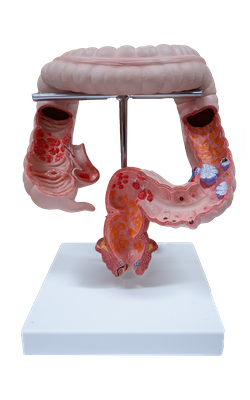
Main Model
Polyp
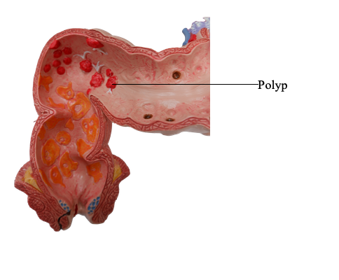
Colonic Polyps and Neoplastic Disease
Polyps are most common in the colon but may occur in the
esophagus, stomach, or small intestine. Those without
stalks are referred to as sessile. As sessile polyps enlarge,
proliferation of cells adjacent to the polyp and the effects
of traction on the luminal protrusion may combine to
create a stalk. Polyps with stalks are termed pedunculated.
In general, intestinal polyps can be classified as nonneoplastic
or neoplastic. The most common neoplastic polyp is the adenoma, which has the potential to progress to cancer.
Nonneoplastic colonic polyps can be further classified as
inflammatory, hamartomatous, or hyperplastic.
Inflammatory Polyps
The solitary rectal ulcer syndrome is associated with a purely
inflammatory polyp. Patients present with the clinical triad of rectal bleeding, mucus discharge, and an inflammatory lesion of the anterior rectal wall. The underlying
cause is impaired relaxation of the anorectal sphincter, creating a sharp angle at the anterior rectal shelf. This leads
to recurrent abrasion and ulceration of the overlying rectal
mucosa. Chronic cycles of injury and healing produce a polypoid mass composed of inflamed and reactive mucosal
tissue.
Hamartomatous Polyps
Hamartomatous polyps occur sporadically and as components of various genetically determined or acquired syndromes. Hamartomas
are disorganized, tumorlike growths composed of mature cell types normally present at the site at which the polyp
develops. Hamartomatous polyposis syndromes are rare,
but they are important to recognize because of associated
intestinal and extraintestinal manifestations and the need
to screen family members.
Juvenile Polyps
Juvenile polyps are the most common type of hamartomatous polyp. They may be sporadic or syndromic. Sporadic
juvenile polyps are usually solitary, but the number varies
from 3 to as many as 100 in individuals with the autosomal
dominant syndrome of juvenile polyposis. In adults, the
sporadic form is sometimes also referred to as an inflammatory polyp, particularly when dense inflammatory infiltrates
are present. The vast majority of juvenile polyps occur
in children younger than 5 years of age. Juvenile polyps
characteristically are located in the rectum and most manifest with rectal bleeding. In some cases, prolapse occurs
and the polyp protrudes through the anal sphincter. Dysplasia occurs in a small proportion of (mostly syndrome-associated) juvenile polyps, and the juvenile polyposis
syndrome is associated with an increased risk for development of adenocarcinoma within the colon and at other
sites. Colectomy may be required to limit the
hemorrhage associated with polyp ulceration in juvenile polyposis.
Morphology
Individual sporadic and syndromic juvenile polyps often are indistinguishable. They typically are pedunculated, smooth-surfaced,
reddish lesions that are less than 3 cm in diameter and display characteristic cystic spaces on cut sections. Microscopic examination shows the spaces to be dilated glands filled with mucin
and inflammatory debris. Some data suggest that
mucosal hyperplasia is the initiating event in polyp development,
and this mechanism is consistent with the discovery that mutations in pathways that regulate cellular growth, such as transforming growth factor-β (TGF-β) signaling, are associated with
autosomal dominant juvenile polyposis.
Peutz-Jeghers Syndrome
Peutz-Jeghers syndrome is a rare autosomal dominant
disorder defined by the presence of multiple gastrointestinal hamartomatous polyps and mucocutaneous
hyperpigmentation that carries an increased risk for development of several malignancies, including cancers
of the colon, pancreas, breast, lung, ovaries, uterus, and
testes, as well as other unusual neoplasms. Germ line loss-of-function mutations in the LKB1/STK11 gene are present
in approximately half of the patients with the familial form
of Peutz-Jeghers syndrome, as well as a subset of patients with the sporadic form. LKB1/STK11 encodes a tumor suppressive protein kinase that regulates cellular metabolism,
yet another example of links between alterned metabolism,
abnormal cell growth, and cancer risk. Intestinal polyps are
most common in the small intestine, although they may also
occur in the stomach and colon and, rarely, in the bladder
and lungs. On gross evaluation, the polyps are large and
pedunculated with a lobulated contour. Histologic examination demonstrates a characteristic arborizing network
of connective tissue, smooth muscle, lamina propria, and
glands lined by normal-appearing intestinal epithelium.
Hyperplastic Polyps
Colonic hyperplastic polyps are common epithelial proliferations that typically are discovered in the sixth and
seventh decades of life. The pathogenesis of hyperplastic
polyps is incompletely understood, but formation of these
lesions is thought to result from decreased epithelial cell turnover and delayed shedding of surface epithelial cells,
leading to a "pileup" of goblet cells. Although these lesions
have no malignant potential, they must be distinguished
from sessile serrated adenomas, histologically similar
lesions that have malignant potential.
Morphology
Hyperplastic polyps are most commonly found in the left colon
and typically are less than 5 mm in diameter.They are smooth,
nodular protrusions of the mucosa, often on the crests of mucosal folds. They may occur singly but more frequently are
multiple, particularly in the sigmoid colon and rectum. Histologically, hyperplastic polyps are composed of mature goblet and
absorptive cells. The delayed shedding of these cells leads to
crowding that creates the serrated surface architecture, the
morphologic hallmark of these lesions.
Adenomas
The most common and clinically important neoplastic
polyps are colonic adenomas, benign polyps that give
rise to a majority of colorectal adenocarcinomas. Most
adenomas, however, do not progress to adenocarcinoma.
Colorectal adenomas are characterized by the presence of
epithelial dysplasia. These growths range from small, often
pedunculated polyps to large sessile lesions. There is no
gender predilection, and they are present in nearly 50% of adults living in the Western world beginning at age 50.
Because these polyps are precursors to colorectal cancer,
current recommendations are that all adults in the United
States undergo screening colonoscopy starting at 50 years
of age. Because individuals with a family history are at risk
for developing colon cancer earlier in life, they are typically screened at least 10 years before the youngest age at
which a relative was diagnosed. While adenomas are less common in Asia, their frequency has risen (in parallel with
an increasing incidence of colorectal adenocarcinoma) as
Western diets and lifestyles become more common.
Morphology
Typical adenomas range from 0.3 to 10 cm in diameter and can be
pedunculated or sessile, with the surface of both
types having a texture resembling velvet or a raspberry, due to the abnormal epithelial growth pattern. Histologically,the cytologic hallmark of epithelial dysplasia is nuclear hyperchromasia, elongation, and stratification. These
changes are most easily appreciated at the surface of the adenoma,
because the epithelium fails to mature as cells migrate out of
the crypt. Pedunculated adenomas have slender fibromuscular
stalks containing prominent blood vessels derived
from the submucosa. The stalk usually is covered by nonneoplastic
epithelium, but dysplastic epithelium is sometimes present.
Adenomas can be classified as tubular, tubulovillous, or
villous on the basis of their architecture. These categories,
however, have little clinical significance in isolation. Tubular adenomas tend to be small, pedunculated polyps composed of
small, rounded, or tubular glands. By contrast, villous
adenomas, which often are larger and sessile, are covered by
slender villi. Tubulovillous adenomas have a mixture
of tubular and villous elements. Although foci of invasion are
more frequent in villous adenomas than in tubular adenomas, villous architecture alone does not increase cancer risk when
polyp size is considered.
The histologic features of sessile serrated adenomas,
which are also referred to as sessile serrated polyps, overlap with
those of hyperplastic polyps and lack typical cytologic features
of dysplasia. Nevertheless, sessile serrated adenomas, which are most common in the right colon, have a malignant
potential similar to that of conventional adenomas. The most
useful histologic feature that distinguishes sessile serrated adenomas from hyperplastic polyps is the presence of serrated architecture throughout the full length of the glands, including the
crypt base, associated with crypt dilation and lateral growth, in
the former. By contrast, serrated architecture typically is confined to the surface of hyperplastic polyps.
Although most colorectal adenomas behave in a benign
fashion, a small proportion harbor invasive cancer at the time of
detection. Size is the most important characteristic that correlates with risk for malignancy. For example, while
cancer is extremely rare in adenomas less than 1 cm in diameter,
some studies suggest that nearly 40% of lesions larger than 4 cm in diameter contain foci of invasive cancer. In addition to size,
high-grade dysplasia is a risk factor for cancer in an individual
polyp (but not other polyps in the same patient).
Familial Syndromes
Several syndromes associated with colonic polyps and
increased rates of colon cancer have been described. The
genetic basis of these disorders has been established and
has greatly enhanced the current understanding of sporadic colon cancer (Table 15.7).
Familial Adenomatous Polyps
Familial adenomatous polyposis (FAP) is an autosomal
dominant disorder marked by the appearance of numerous colorectal adenomas by the teenage years. It is caused
by mutations of the adenomatous polyposis coli gene (APC).
A count of at least 100 polyps is necessary for a diagnosis of
classic FAP, and as many as several thousand may be
present (Fig. 15.35). Except for their remarkable numbers,
these growths are morphologically indistinguishable from
sporadic adenomas. Colorectal adenocarcinoma develops
in 100% of patients with untreated FAP, often before 30
years of age. As a result, prophylactic colectomy is standard therapy for individuals carrying APC mutations.
However, patients remain at risk for extraintestinal manifestations, including neoplasia at other sites. Specific APC
mutations are also associated with the development of
other manifestations of FAP and explain variants such as
Gardner syndrome and Turcot syndrome. In addition to intestinal polyps, clinical features of Gardner syndrome, a
variant of FAP, may include osteomas of the mandible,
skull, and long bones; epidermal cysts; desmoid and
thyroid tumors; and dental abnormalities, including
unerupted and supernumerary teeth. Turcot syndrome is
rarer and is characterized by intestinal adenomas and
tumors of the central nervous system. Two-thirds of patients with Turcot syndrome have APC gene mutations
and develop medulloblastomas. The remaining one-third
have mutations in one of several genes involved in DNA
repair and develop glioblastomas. Some patients with hundreds of adenomas lack APC mutations but instead have
mutations of the base excision repair gene MUTYH (also
called MUTYH polyposis). The role of these genes in tumor
development is discussed later.
Hereditary Nonpolyposis Colorectal Cancer
Hereditary nonpolyposis colorectal cancer (HNPCC), also
known as Lynch syndrome, originally was described as
familial clustering of cancers at several sites including the
colorectum, endometrium, stomach, ovary, ureters, brain, small bowel, hepatobiliary tract, and skin. Colon cancers
in patients with HNPCC tend to occur at younger ages
than do sporadic colon cancers and often are located in the
right colon (Table 15.7). Adenomas are present in HNPCC,
but excessive numbers (i.e., polyposis) is not. In many
cases, sessile serrated adenomas are associated with
HNPCC, and mucin production may be a prominent in the
subsequent adenocarcinomas.
Just as identification of APC mutations in FAP has provided molecular insights into the pathogenesis of a majority of sporadic colon cancers, dissection of the defects in
HNPCC has shed light on the mechanisms responsible for
most of the remaining sporadic cases. HNPCC is caused
by inherited germ line mutations in genes that encode
proteins responsible for the detection, excision, and
repair of errors that occur during DNA replication. At
least five such mismatch repair genes have been recognized, but a majority of HNPCC cases involve either MSH2
or MLH1. Patients with HNPCC inherit one mutated DNA
repair gene and one normal allele. When the second copy
is lost through mutation or epigenetic silencing, defects in
mismatch repair lead to the accumulation of mutations at
rates up to 1000 times higher than normal, mostly in regions
containing short repeating DNA sequences referred to as
microsatellite DNA. The human genome contains approximately 50,000 to 100,000 microsatellites, which are prone
to undergo expansion during DNA replication and represent the most frequent sites of mutations in HNPCC. The
consequences of mismatch repair defects and the resulting
microsatellite instability are discussed next in the context of
colonic adenocarcinoma.
SUMMARY
COLONIC POLYPS, ADENOMAS, AND
ADENOCARCINOMAS
• Intestinal polyps can be classified as nonneoplastic or neoplastic.
The nonneoplastic polyps can be further defined as inflammatory, hamartomatous, or hyperplastic.
• Inflammatory polyps form as a result of chronic cycles of injury
and healing.
• Hamartomatous polyps occur sporadically or as a part of genetic
diseases. In the latter case, they often are associated with
increased risk for malignancy.
• Hyperplastic polyps are benign epithelial proliferations most
commonly found in the left colon and rectum. They are not
reactive in origin, in contrast with gastric hyperplastic polyps;
have no malignant potential; and must be distinguished from
sessile serrated adenomas or polyps.
• Benign epithelial neoplastic polyps of the colon are termed adenomas.The hallmark feature of these lesions, which are the precursors of colonic adenocarcinomas, is cytologic dysplasia.
• In contrast with traditional adenomas,sessile serrated adenomas,
or polyps, lack cytologic dysplasia and share some morphologic
features with hyperplastic polyps.
• Familial adenomatous polyposis (FAP) and hereditary nonpolyposis
colorectal cancer (HNPCC) are the most common forms of
familial colon cancer. FAP is caused by APC mutations, and
patients typically have over 100 adenomas and develop colon
cancer before 30 years of age.
• HNPCC is caused by mutations in DNA mismatch repair genes.
Patients with HNPCC have far fewer polyps and develop cancer
at an older age than that typical for patients with FAP but at a
younger age than in patients with sporadic colon cancer.