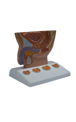
Main Model
Coccygeal bone
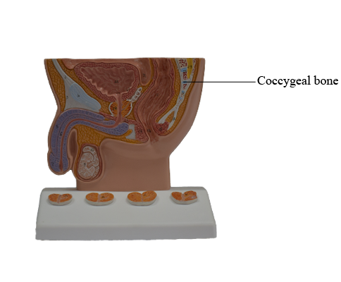
Coccyx
The coccyx (tail bone) is a small triangular bone that is usually formed by fusion of the four rudimentary coccygeal vertebrae, although in some people, there may be one less or
one more. Coccygeal vertebra 1 (Co1)
may remain separate from the fused group. The coccyx is the remnant of the skeleton of the embryonic tail-like caudal
eminence, which is present in human embryos from the end
of the 4th week until the beginning of the 8th week. The pelvic surface of the coccyx
is concave and relatively smooth, and the posterior surface
has rudimentary articular processes. Co1 is the largest and
broadest of all the coccygeal vertebrae. Its short transverse
processes are connected to the sacrum, and its rudimentary articular processes form coccygeal cornua, which articulate
with the sacral cornua. The last three coccygeal vertebrae
often fuse during middle life, forming a beak-like coccyx; this
accounts for its name (Greek coccyx, cuckoo). With increasing
age, Co1 often fuses with the sacrum, and the remaining coccygeal vertebrae usually fuse to form a single bone.
The coccyx does not participate with the other vertebrae
in support of the body weight when standing; however, when
sitting it may flex anteriorly somewhat, indicating that it is
receiving some weight. The coccyx provides attachments for
parts of the gluteus maximus and coccygeus muscles and the
anococcygeal ligament, the median fibrous band of the pubococcygeus muscles.