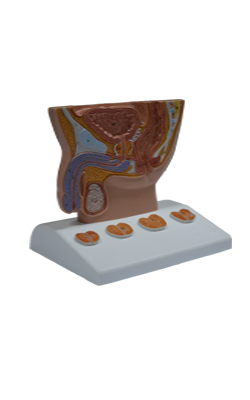
Main Model
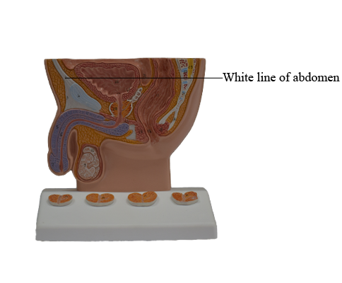
White line of abdomen
Rectus Sheath, Linea Alba, and Umbilical Ring
The rectus sheath is the strong, incomplete fibrous compartment of the rectus abdominis and pyramidalis muscles. Also found in the rectus sheath are the superior
and inferior epigastric arteries and veins, lymphatic vessels,
and distal portions of the thoraco-abdominal nerves (abdominal portions of the anterior rami of spinal nerves T7-T12).
The rectus sheath is formed by the decussation and interweaving of the aponeuroses of the flat abdominal muscles. The external oblique aponeurosis contributes to
the anterior wall of the sheath throughout its length. The
superior two thirds of the internal oblique aponeurosis splits into two layers (laminae) at the lateral border of the rectus
abdominis; one lamina passing anterior to the muscle and the other passing posterior to it. The anterior lamina joins the aponeurosis of the external oblique to form the anterior layer
of the rectus sheath. The posterior lamina joins the aponeurosis of the transversus abdominis to form the posterior layer of the rectus sheath.
Beginning approximately one third of the distance from
the umbilicus to the pubic crest, the aponeuroses of the three
flat muscles pass anterior to the rectus abdominis to form
the anterior layer of the rectus sheath, leaving only the relatively thin transversalis fascia to cover the rectus abdominis
posteriorly. A crescentic arcuate line demarcates
the transition between the aponeurotic posterior wall of the
sheath covering the superior three quarters of the rectus and the transversalis fascia covering the inferior quarter.
Throughout the length of the sheath, the fibers of the anterior and posterior layers of the sheath interlace in the anterior median line to form the complex linea alba (Latin white line).
The posterior layer of the rectus sheath is also deficient
superior to the costal margin because the transversus abdominis is continued superiorly as the transversus thoracis, which
lies internal to the costal cartilages, and
the internal oblique attaches to the costal margin. Hence, superior to the costal margin, the rectus abdominis lies
directly on the thoracic wall.
The linea alba, running vertically the length of the anterior
abdominal wall and separating the bilateral rectus sheaths, narrows inferior to the umbilicus to the width
of the pubic symphysis and widens superiorly to the width
of the xiphoid process. The linea alba transmits small vessels
and nerves to the skin. In thin muscular people, a groove
is visible in the skin overlying the linea alba. At its middle,
underlying the umbilicus, the linea alba contains the umbilical ring, a defect in the linea alba through which the fetal
umbilical vessels passed to and from the umbilical cord and
placenta. All layers of the anterolateral abdominal wall fuse
at the umbilicus. As fat accumulates in the subcutaneous tissue postnatally, the skin becomes raised around the umbilical
ring and the umbilicus becomes depressed. This occurs 7-14
days after birth, when the atrophic umbilical cord "falls off".