Main Model
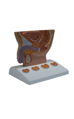
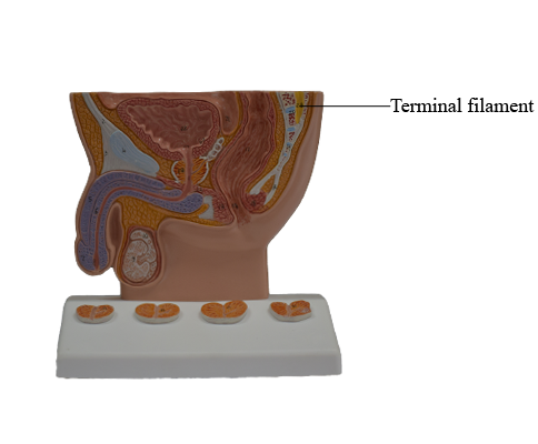
Terminal filament
Spinal Cord
The spinal cord is the major reflex center and conduction
pathway between the body and brain. This cylindrical structure, slightly flattened anteriorly and posteriorly, is protected
by the vertebrae, their associated ligaments and muscles, the
spinal meninges, and the cerebrospinal fluid (CSF).
The spinal cord begins as a continuation of the medulla
oblongata (often called the medulla), the caudal part of the
brainstem. In adults, the spinal cord
is 42-45 cm long and extends from the foramen magnum
in the occipital bone to the level of the L1 or L2 vertebra. However, its tapering inferior end, the conus
medullaris, may terminate as high as T12 vertebra or as low
as L3 vertebra. Thus the spinal cord occupies only the superior two thirds of the vertebral canal.
The spinal cord is enlarged in two regions in relationship
to innervation of the limbs. The cervical enlargement
extends from C4 through T1 segments of the spinal cord,
and most of the anterior rami of the spinal nerves arising
from it form the brachial plexus of nerves that innervates the
upper limbs. The lumbosacral enlargement extends from
T11 through S1 segments of the spinal cord, inferior to which
the cord continues to diminish as the conus medullaris. The
anterior rami of the spinal nerves arising from this enlargement make up the lumbar and sacral plexuses of nerves that
innervate the lower limbs.
Spinal Nerves and Nerve Roots
The portion of the spinal cord giving rise to the
rootlets and roots that ultimately form one bilateral pair of
spinal nerves is designated a spinal cord segment, the identity of which is the same as the spinal nerves arising from it.
Cervical spinal nerves (except C8) bear the same alphanumeric designation as the vertebrae forming the inferior
margin of the IV foramina through which the nerve exits the
vertebral canal. The more inferior spinal (T1 through Co1)
nerves bear the same alphanumeric designation as the vertebrae forming the superior margin of their exit. First cervical nerves lack posterior roots in 50% of people, and
the coccygeal nerve may be absent.
In embryos, the spinal cord occupies the whole length of
the vertebral canal; thus cord segments lie approximately at the vertebral level of the same number, the spinal
nerves passing laterally to exit the corresponding IV foramen.
By the end of the embryonic period (8th week), the tail-like
caudal eminence has disappeared, and the number of coccygeal vertebrae is reduced from six to four segments. The
spinal cord in the vertebral canal of the coccyx atrophies.
During the fetal period, the vertebral column grows faster
than the spinal cord; as a result, the cord "ascends" relative
to the vertebral canal. At birth, the tip of the conus medullaris is at the L4-L5 level. Thus, in postnatal life, the spinal cord is shorter than the vertebral column; consequently,
there is a progressive obliquity of the spinal nerve roots. Because the distance between the origin of a
nerve's roots from the spinal cord and the nerve's exit from
the vertebral canal increases as the inferior end of the vertebral column is approached, the length of the nerve roots also
increases progressively.
The lumbar and sacral nerve roots are therefore the longest, extending far beyond the termination of the adult spinal
cord at approximately the L2 level to reach the remaining lumbar, sacral, and coccygeal IV foramina. This loose bundle
of spinal nerve roots, arising from the lumbosacral enlargement and the conus medullaris and coursing within the lumbar cistern of CSF caudal to the termination of the spinal
cord, resembles a horse's tail, hence its name - the cauda
equina (Latin horse tail).
Arising from the tip of the conus medullaris, the filum terminale descends among the spinal nerve roots in the cauda
equina. The filum terminale is the vestigial remnant of the
caudal part of the spinal cord that was in the tail-like caudal
eminence of the embryo. Its proximal end (the filum terminale internum, or pial part of the terminal filum) consists
of vestiges of neural tissue, connective tissue, and neuroglial
tissue covered by pia mater. The filum terminale perforates
the inferior end of the dural sac, gaining a layer of dura and
continuing through the sacral hiatus as the filum terminale externum (or dural part of the terminal filum, also known as
the coccygeal ligament) to attach to the dorsum of the coccyx.
The filum terminale is an anchor for the inferior end of the
spinal cord and spinal meninges.