Main Model
Sacral bone
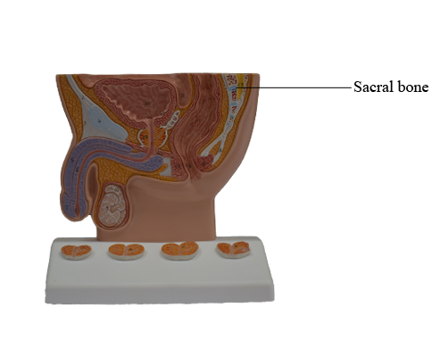
Sacrum
The wedged-shaped sacrum (Latin sacred bone) is usually
composed of five fused sacral vertebrae in adults.
It is located between the hip bones and forms the roof and
posterosuperior wall of the posterior half of the pelvic cavity. The triangular shape of the sacrum results from the
rapid decrease in the size of the inferior lateral masses of
the sacral vertebrae during development. The inferior half
of the sacrum is not weight-bearing; therefore, its bulk is diminished considerably. The sacrum provides strength and
stability to the pelvis and transmits the weight of the body to
the pelvic girdle, the bony ring formed by the hip bones and
sacrum, to which the lower limbs are attached.
The sacral canal is the continuation of the vertebral canal
in the sacrum. It contains the bundle of spinal
nerve roots arising inferior to the L1 vertebra, known as the
cauda equina (Latin horse tail), that descend past the termination of the spinal cord. On the pelvic and posterior surfaces of
the sacrum between its vertebral components are typically four
pairs of sacral foramina for the exit of the posterior and anterior rami of the spinal nerves. The anterior (pelvic) sacral foramina are larger than the posterior (dorsal) ones.
The base of the sacrum is formed by the superior surface of the S1 vertebra. Its superior articular
processes articulate with the inferior articular processes of
the L5 vertebra. The anterior projecting edge of the body
of the S1 vertebra is the sacral promontory (Latin mountain ridge), an important obstetrical landmark.
The apex of the sacrum, its tapering inferior end, has an
oval facet for articulation with the coccyx.
The sacrum supports the vertebral column and forms the
posterior part of the bony pelvis. The sacrum is tilted so that
it articulates with the L5 vertebra at the lumbosacral angle, which varies from 130° to 160°. The sacrum is
often wider in proportion to length in the female than in the male, but the body of the S1 vertebra is usually larger in males.
The pelvic surface of the sacrum is smooth and concave. Four transverse lines on this surface of
sacra from adults indicate where fusion of the sacral vertebrae occurred. During childhood, the individual sacral vertebrae are connected by hyaline cartilage and separated by IV
discs. Fusion of the sacral vertebrae starts after age 20; however, most of the IV discs remain unossified up to or beyond
middle life.
The dorsal surface of the sacrum is rough, convex, and
marked by five prominent longitudinal ridges.
The central ridge, the median sacral crest, represents the
fused rudimentary spinous processes of the superior three
or four sacral vertebra; S5 has no spinous process. The intermediate sacral crests represent the fused articular
processes, and the lateral sacral crests are the tips of the
transverse processes of the fused sacral vertebrae.
The clinically important features of the dorsal surface of
the sacrum are the inverted U-shaped sacral hiatus and the
sacral cornua (Latin horns). The sacral hiatus results from the
absence of the laminae and spinous process of S5 and sometimes S4. The sacral hiatus leads into the sacral canal. Its
depth varies, depending on how much of the spinous process
and laminae of S4 are present. The sacral cornua, representing the inferior articular processes of S5 vertebra, project
inferiorly on each side of the sacral hiatus and are a helpful
guide to its location.
The superior part of the lateral surface of the sacrum
looks somewhat like an auricle (Latin external ear); because
of its shape, this area is called the auricular surface. It is the site of the synovial part of the sacro-iliac
joint between the sacrum and ilium. During life, the auricular surface is covered with hyaline cartilage.