Main Model
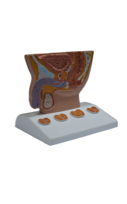
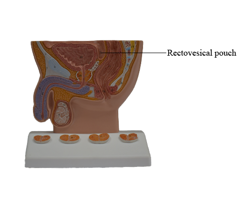
Rectovesical pouch
Peritoneum and Peritoneal Cavity of Pelvis
The parietal peritoneum lining the abdominal cavity continues inferiorly into the pelvic cavity, but does not reach
the pelvic floor. Instead, it reflects onto the pelvic viscera,
remaining separated from the pelvic floor by the pelvic viscera and the surrounding pelvic fascia. Except for
the ovaries and uterine tubes, the pelvic viscera are not completely ensheathed by the peritoneum, lying inferior to it for
the main part. Only their superior and superolateral surfaces are covered with peritoneum. Only the uterine tubes (except
for their ostia, which are open) are intraperitoneal and suspended by a mesentery. The ovaries, although suspended in
the peritoneal cavity by a mesentery, are not covered with
glistening peritoneum; instead a special, relatively dull epithelium of cuboidal cells (germinal epithelium) covers them.
A loose areolar (fatty) layer between the transversalis fascia
and the parietal peritoneum of the inferior part of the anterior abdominal wall allows the bladder to expand between
these layers as it becomes distended with urine. The region
superior to the bladder is the only site where the parietal peritoneum is not firmly bound to the underlying
structures. Consequently, the level at which the peritoneum
reflects onto the superior surface of the bladder, creating the supravesical fossa, is variable, depending
on the fullness of the bladder. When the peritoneum reflects
from the abdominopelvic wall onto the pelvic viscera and fascia, a series of folds and fossae is created.
In the female, as the peritoneum at or near the midline
reaches the posterior border of the roof of the bladder, it
reflects onto the anterior aspect of the uterus at the isthmus
of the uterus; thus it is not related to the anterior vaginal fornix, which is
subperitoneal in location. The peritoneum passes over the
fundus of the uterus and descends the entire posterior aspect
of the uterus onto the posterior vaginal wall before reflecting
superiorly onto the anterior wall of the inferior rectum (rectal
ampulla). The "pocket" thus formed between the uterus and
the rectum is the recto-uterine pouch (cul-de-sac of Douglas). The median recto-uterine pouch is often
described as being the inferiormost extent of the peritoneal
cavity in the female, but often its lateral extensions on each
side of the rectum, the pararectal fossae, are deeper.
Prominent peritoneal ridges, the recto-uterine folds,
formed by underlying fascial ligaments demarcate the lateral
boundaries of the pararectal fossae. As the peritoneum passes up and over the uterus in the middle of the
pelvic cavity, a double peritoneal fold, the broad ligament of
the uterus, extends between the uterus and the lateral pelvic
wall on each side, forming a partition that separates the paravesical fossae and pararectal fossae of each side. The uterine tubes, ovaries, ligaments of the ovaries, and round ligaments
of the uterus are enclosed within the broad ligaments. Recall that in females the pelvic peritoneal cavity communicates with the external environment via the uterine tubes, uterus, and vagina.
In males and in females who have had a hysterectomy
(removal of the uterus), the central peritoneum descends a
short distance (as much as 2 cm) down the posterior surface
(base) of the bladder, and then reflects superiorly onto the
anterior surface of the inferior rectum, forming the rectovesical pouch. The female recto-uterine pouch is normally deeper
(extends farther caudally) than the male rectovesical pouch.
In the male, a gentle peritoneal fold or ridge, the ureteric
fold, is formed as the peritoneum passes up and over the ureter and ductus (vas) deferens (secretory duct of the testis) on
each side of the posterior bladder, separating the paravesical
and pararectal fossae; in this regard, it is the male equivalent of
the broad ligament. Posterior to the ureteric folds and lateral to
the central rectovesical pouch, the peritoneum often descends
far enough caudally to cover the superior ends or superior posterior surfaces of the seminal glands (vesicles) and ampullae of the ductus deferens. Except for these sites (and the testis in its
tunica vaginalis, which is derived from peritoneum), the male
reproductive organs are not in contact with the peritoneum.
In both sexes, the inferior third of the rectum is below the
inferior limits of the peritoneum (i.e., it is subperitoneal), the
middle third is covered with peritoneum only on its anterior surface, and the superior third is covered on both its anterior
and its lateral surfaces. The rectosigmoid junction, near the
pelvic brim, is intraperitoneal.