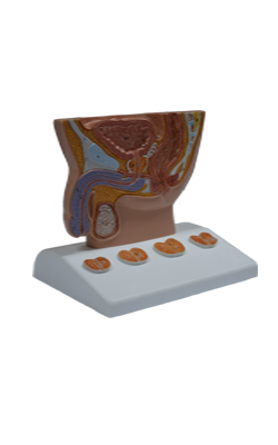
Main Model
Testicle
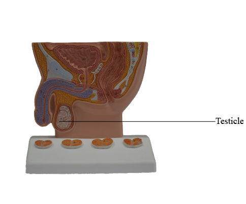
Testes
The testes (testicles) are the male gonads - paired ovoid reproductive glands that produce sperms (spermatozoa) and male hormones, primarily testosterone. The testes are suspended in the scrotum by the spermatic cords, with the left testis usually suspended (hanging) more inferiorly than the right testis.
The surface of each testis is covered by the visceral layer of the tunica vaginalis, except where the testis attaches to the epididymis and spermatic cord. The tunica vaginalis is a closed peritoneal sac partially surrounding the testis, which represents the closed-off distal part of the embryonic processus vaginalis. The visceral layer of the tunica vaginalis is closely applied to the testis, epididymis, and inferior part of the ductus deferens. The slit-like recess of the tunica vaginalis, the sinus of the epididymis, is between the body of the epididymis and the posterolateral surface of the testis.
The parietal layer of the tunica vaginalis, adjacent to the internal spermatic fascia, is more extensive than the visceral layer and extends superiorly for a short distance onto the distal part of the spermatic cord. The small amount of fluid in the cavity of the tunica vaginalis separates the visceral and parietal layers, allowing the testis to move freely in the scrotum.
The testes have a tough fibrous outer surface, the tunica albuginea, that thickens into a ridge on its internal, posterior aspect as the mediastinum of the testis. From this internal ridge, fibrous septa extend inward between lobules of minute but long and highly coiled seminiferous tubules in which the sperms are produced. The seminiferous tubules are joined by straight tubules to the rete testis (Latin rete, a net), a network of canals in the mediastinum of the testis.
The long testicular arteries arise from the anterolateral aspect of the abdominal aorta just inferior to the renal arteries. They pass retroperitoneally (posterior to the peritoneum) in an oblique direction, crossing over the ureters and the inferior parts of the external iliac arteries to reach the deep inguinal rings. They enter the inguinal canals through the deep rings, pass through the canals, exit them through the superficial inguinal rings, and enter the spermatic cords to supply the testes. The testicular artery or one of its branches anastomoses with the artery of the ductus deferens.
The veins emerging from the testis and epididymis form the pampiniform venous plexus, a network of 8-12 veins lying anterior to the ductus deferens and surrounding the testicular artery in the spermatic cord. The pampiniform plexus is part of the thermoregulatory system of the testis (along with the cremasteric and dartos muscles) helping to keep this gland at a constant temperature. The veins of each pampiniform plexus converge superiorly, forming a right testicular vein, which enters the inferior vena cava (IVC), and a left testicular vein, which enters the left renal vein.
The lymphatic drainage of the testis follows the testicular artery and vein to the right and left lumbar (caval/ aortic) and pre-aortic lymph nodes.
The autonomic nerves of the testis arise as the testicular plexus of nerves on the testicular artery, which contains vagal parasympathetic and visceral afferent fibers and sympathetic fibers from the T10(-T11) segment of the spinal cord.