Main Model
1 Kidney
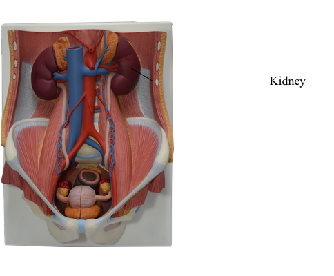
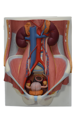
The ovoid kidneys remove excess water, salts, and wastes of protein metabolism from the blood while returning nutrients and chemicals to the blood. They lie retroperitoneally on the posterior abdominal wall, one on each side of the vertebral column at the level of the T12-L3 vertebrae.
At the concave medial margin of each kidney is a vertical cleft, the renal hilum. The renal hilum is the entrance to a space within the kidney, the renal sinus. Structures that serve the kidneys (vessels, nerves, and structures that drain urine from the kidney) enter and exit the renal sinus through the renal hilum. The hilum of the left kidney lies near the transpyloric plane, approximately 5 cm from th median plane. The transpyloric plane passes through the superior pole of the right kidney, which is approximately 2.5 cm lower than the left pole, probably due to the presence of the liver. Posteriorly, the superior parts of the kidneys lie deep to the 11th and 12th ribs. The levels of the kidneys change during respiration and with changes in posture. Each kidney moves 2-3 cm in a vertical direction during the movement of the diaphragm that occurs with deep breathing. Because the usual surgical approach to the kidneys is through the posterior abdominal wall, it is helpful to know that the inferior pole of the right kidney is approximately a finger’s breadth superior to the iliac crest.
During life, the kidneys are reddish brown and measure approximately 10 cm in length, 5 cm in width, and 2.5 cm in thickness. Superiorly, the posterior aspects of the kidneys are associated with the diaphragm, which separates them from the pleural cavities and the 12th pair of ribs. More inferiorly, the posterior surfaces of the kidney are related to the psoas major muscles medially and the quadratus lumborum muscle. The subcostal nerve and vessels and the iliohypogastric and ilio-inguinal nerves descend diagonally across the posterior surfaces of the kidneys. The liver, duodenum, and ascending colon are anterior to the right kidney. This kidney is separated from the liver by the hepatorenal recess. The left kidney is related to the stomach, spleen, pancreas, jejunum, and descending colon.
At the hilum, the renal vein is anterior to the renal artery, which is anterior to the renal pelvis. Within the kidney, the renal sinus is occupied by the renal pelvis, calices, vessels, and nerves, and a variable amount of fat. Each kidney has anterior and posterior surfaces, medial and lateral margins, and superior and inferior poles. However, because of the protrusion of the lumbar vertebral column into the abdominal cavity, the kidneys are obliquely placed, lying at an angle to each other. Consequently, the transverse diameter of the kidneys is foreshortened in anterior views and anteroposterior (AP) radiographs. The lateral margin of each kidney is convex, and the medial margin is concave where the renal sinus and renal pelvis are located. The indented medial margin gives the kidney a somewhat bean-shaped appearance.
The renal pelvis is the flattened, funnel-shaped expansion of the superior end of the ureter. The apex of the renal pelvis is continuous with the ureter. The renal pelvis receives two or three major calices (calyces), each of which divides into two or three minor calices. Each minor calyx is indented by a renal papilla, the apex of the renal pyramid, from which the urine is excreted. In living persons, the renal pelvis and its calices are usually collapsed (empty). The pyramids and their associated cortex form the lobes of the kidney. The lobes are visible on the external surfaces of the kidneys in fetuses, and evidence of the lobes may persist for some time after birth.