Main Model
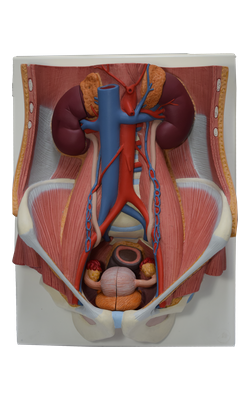
22 Transversus abdominis
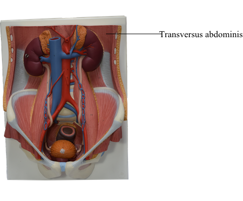
The fibers of the transversus abdominis, the innermost of the three flat abdominal muscles, run more or less transversally, except for the inferior ones, which run parallel to those of the internal oblique. This transverse, circumferential orientation is ideal for compressing the abdominal contents, increasing intra-abdominal pressure. The fibers of the transversus abdominis muscle also end in an aponeurosis, which contributes to the formation of the rectus sheath.
The attachments of the transversus abdominis are:
Origin: Internal surfaces of 7th-12th costal cartilages, thoracolumbar fascia, iliac crest, and connective tissue deep to lateral third of inguinal ligament
Insertion: Linea alba with aponeurosis of internal oblique, pubic crest, and pecten pubis via conjoint tendon
Innervation: Thoraco-abdominal nerves (anterior rami of T6-T12 spinal nerves) and first lumbar nerves
Main Action(s): Compresses and supports abdominal viscera
Between the internal oblique and the transversus abdominis muscles is a neurovascular plane, which corresponds with a similar plane in the intercostal spaces. In both regions, the plane lies between the middle and deepest layers of muscle. The neurovascular plane of the anterolateral abdominal wall contains the nerves and arteries supplying the anterolateral abdominal wall. In the anterior part of the abdominal wall, the nerves and vessels leave the neurovascular plane and lie mostly in the subcutaneous tissue.