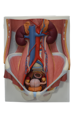
Main Model
10 Suprarenal gland
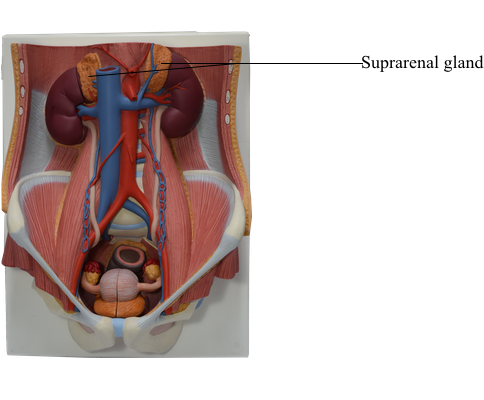
The suprarenal (adrenal) glands, yellowish in living persons, are located between the superomedial aspects of the kidneys and the diaphragm, where they are surrounded by connective tissue containing considerable perinephric fat. The suprarenal glands are enclosed by renal fascia by which they are attached to the crura of the diaphragm. Although the name “suprarenal” implies that the kidneys are their primary relationship, their major attachment is to the diaphragmatic crura. They are separated from the kidneys by a thin septum (part of the renal fascia).
The shape and relations of the suprarenal glands differ on the two sides. The pyramidal right gland is more apical (situated over the superior pole) relative to the left kidney, lies anterolateral to the right crus of the diaphragm, and makes contact with the inferior vena cava (IVC) anteromedially and the liver anterolaterally. The crescent-shaped left gland is medial to the superior half of the left kidney and is related to the spleen, stomach, pancreas, and the left crus of the diaphragm.
Each gland has a hilum, where the veins and lymphatic vessels exit the gland, whereas, the arteries and nerves enter the glands at multiple sites. The medial borders of the suprarenal glands are 4-5 cm apart. In this area, from right to left, are the inferior vena cava (IVC), right crus of the diaphragm, celiac ganglion, celiac trunk, superior mesenteric artery (SMA), and the left crus of the diaphragm.
Each suprarenal gland has two parts: the suprarenal cortex and suprarenal medulla; these parts have different embryological origins and different functions.
The suprarenal cortex derives from mesoderm and secretes corticosteroids and androgens. These hormones cause the kidneys to retain sodium and water in response to stress, increasing the blood volume and blood pressure. They also affect muscles and organs such as the heart and lungs.
The suprarenal medulla is a mass of nervous tissue permeated with capillaries and sinusoids that derives from neural crest cells associated with the sympathetic nervous system. The chromaffin cells of the medulla are related to sympathetic ganglion (postsynaptic) neurons in both derivation (neural crest cells) and function. These cells secrete catecholamines (mostly epinephrine) into the bloodstream in response to signals from presynaptic neurons. Powerful medullary hormones, epinephrine (adrenaline) and norepinephrine (noradrenaline), activate the body to a flight-or-fight status in response to traumatic stress. They also increase heart rate and blood pressure, dilate the bronchioles, and change blood flow patterns, preparing for physical exertion.
The endocrine function of the suprarenal glands makes their abundant blood supply necessary. The suprarenal arteries branch freely before entering each gland so that 50-60 branches penetrate the capsule covering the entire surface of the glands. Suprarenal arteries arise from three sources:
• Superior suprarenal arteries (6-8) from the inferior phrenic arteries.
• Middle suprarenal arteries (1) from the abdominal aorta near the level of origin of the superior mesenteric artery (SMA).
• Inferior suprarenal arteries (1) from the renal arteries.
The venous drainage of the suprarenal glands occurs via large suprarenal veins. The short right suprarenal vein drains into the inferior vena cava (IVC), whereas the longer left suprarenal vein, often joined by the inferior phrenic vein, empties into the left renal vein.
The suprarenal lymphatic vessels arise from a plexus deep to the capsule of the gland and from one in its medulla. The lymph passes to the lumbar lymph nodes. Many lymphatic vessels leave the suprarenal glands.
The rich nerve supply of the suprarenal glands is from the celiac plexus and abdominopelvic (greater, lesser, and least) splanchnic nerves. Myelinated presynaptic sympathetic fibers - mainly derived from the intermediolateral cell column (IML), or lateral horn, of gray matter of the spinal cord segments T10-L1 - traverse both the paravertebral and the prevertebral ganglia, without synapse, to be distributed to the chromaffin cells in the suprarenal medulla.