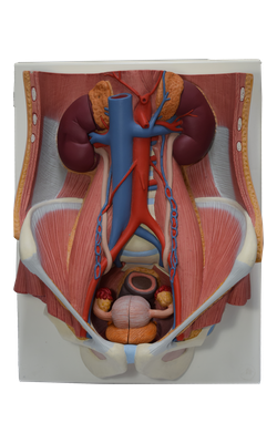
Main Model
8 Urethra
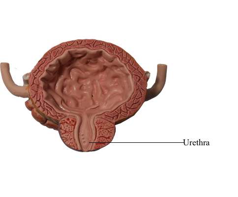
Proximal (Pelvic) Male Urethra
The male urethra is a muscular tube (18-22 cm long) that conveys urine from the internal urethral orifice of the urinary bladder to the external urethral orifice, located at the tip of the glans penis in males. The urethra also provides an exit for semen (sperms and glandular secretions). For descriptive purposes, the urethra is divided into four parts.
The intramural (preprostatic) part of the urethra varies in diameter and length, depending on whether the bladder is filling (bladder neck is tonically contracted so the internal urethral orifice is small and high; the filling internal urethral orifice) or emptying (the neck is relaxed so the orifice is wide and low; the emptying internal urethral orifice). The most prominent feature of the prostatic urethra is the urethral crest, a median ridge between bilateral grooves, the prostatic sinuses. The secretory prostatic ducts open into the prostatic sinuses. The seminal colliculus is a rounded eminence in the middle of the urethral crest with a slit-like orifice that opens into a small cul-de-sac, the prostatic utricle. The utricle is the vestigial remnant of the embryonic uterovaginal canal, the surrounding walls of which, in the female, constitute the primordium of the uterus and a part of the vagina. The ejaculatory ducts open into the prostatic urethra via minute, slit-like openings located adjacent to and occasionally just within the orifice of the prostatic utricle. Thus urinary and reproductive tracts merge at this point.
The intramural and prostatic parts of the urethra are supplied by prostatic branches of the inferior vesical and middle rectal arteries. The veins from the proximal two parts of the urethra drain into the prostatic venous plexus.
The nerves are derived from the prostatic plexus (mixed sympathetic, parasympathetic, and visceral afferent fibers). The prostatic plexus is one of the pelvic plexuses (an inferior extension of the vesical plexus) arising as organ-specific extensions of the inferior hypogastric plexus.
Female Urethra
The female urethra (approximately 4 cm long and 6 mm in diameter) passes antero-inferiorly from the internal urethral orifice of the urinary bladder, posterior and then inferior to the pubic symphysis, to the external urethral orifice. The musculature surrounding the internal urethral orifice of the female bladder is not organized into an internal sphincter. The female external urethral orifice is located in the vestibule of the vagina, the cleft between the labia minora of the external genitalia, directly anterior to the vaginal orifice. The urethra lies anterior to the vagina (forming an elevation in the anterior vaginal wall); its axis is parallel to that of the vagina. The urethra passes with the vagina through the pelvic diaphragm, external urethral sphincter, and perineal membrane.
Urethral glands are present, particularly in the superior part of the urethra. One group of glands on each side, the paraurethral glands, are homologs to the prostate. These glands have a common para-urethral duct, which opens (one on each side) near the external urethral orifice. The external urethral sphincter is located in the perineum.
Blood is supplied to the female urethra by the internal pudendal and vaginal arteries. The veins follow the arteries and have similar names.
The nerves to the urethra arise from the vesical (nerve) plexus and the pudendal nerve. The pattern is similar to that in the male, given the absence of a prostatic plexus and an internal urethra sphincter. Visceral afferents from most of the urethra run in the pelvic splanchnic nerves, but the termination receives somatic afferents from the pudendal nerve. Both the visceral and the somatic afferent fibers extend from cell bodies in the S2-S4 spinal ganglia.