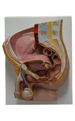
Main Model
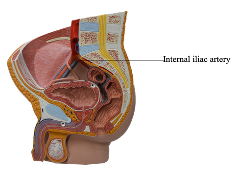
Internal iliac artery
Abdominal Aorta
Most arteries supplying the posterior abdominal wall arise from the abdominal aorta. The subcostal arteries arise from the thoracic aorta and distribute inferior to the 12th rib. The abdominal aorta is approximately 13 cm in length. It begins at the aortic hiatus in the diaphragm at the level of the T12 vertebra and ends at the level of the L4 vertebra by dividing into the right and left common iliac arteries. The abdominal aorta may be represented on the anterior abdominal wall by a band (approximately 2 cm wide) extending from a median point, approximately 2.5 cm superior to the transpyloric plane to a point slightly (2-3 cm) inferior to and to the left of the umbilicus at the level of the supracristal plane (plane of the highest points of the iliac crests). In children and lean adults, the lower abdominal aorta is sufficiently close to the anterior abdominal wall that its pulsations may be detected or apparent when the wall is relaxed.
The common iliac arteries diverge and run inferolaterally, following the medial border of the psoas muscles to the pelvic brim. Here each common iliac artery divides into the internal and external iliac arteries. The internal iliac artery enters the pelvis. The external iliac artery follows the iliopsoas muscle. Just before leaving the abdomen, the external iliac artery gives rise to the inferior epigastric and deep circumflex iliac arteries, which supply the anterolateral abdominal wall.