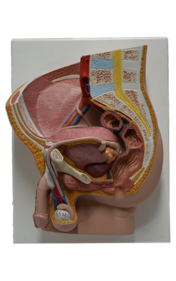
Main Model

Aponeurosis of obliquus externus abdominis muscle

The external oblique muscle is the largest and most superficial of the three flat anterolateral abdominal muscles. In contrast to the two deeper layers, the external oblique does not originate posteriorly from the thoracolumbar fascia; its posterior most fibers (the thickest part of the muscle) have a free edge where they span between its costal origin and the iliac crest. The fleshy part of the muscle contributes primarily to the lateral part of the abdominal wall. Its aponeurosis contributes to the anterior part of the wall.
Although the posteriormost fibers from rib 12 are nearly vertical as they run to the iliac crest, more anterior fibers fan out, taking an increasingly medial direction so that most of the fleshy fibers run inferomedially - in the same direction as the fingers do when the hands are in one's side pockets - with the most anterior and superior fibers approaching a horizontal course. The muscle fibers become aponeurotic approximately at the midclavicular line (MCL) medially and at the spino-umbilical line (line running from the umbilicus to the ASIS) inferiorly, forming a sheet of tendinous fibers that decussate at the linea alba, most becoming continuous with tendinous fibers of the contralateral internal oblique. Thus, the contralateral external and internal oblique muscles together form a “digastric muscle,” a two-bellied muscle sharing a common central tendon that works as a unit. For example, the right external oblique and left internal oblique work together when flexing and rotating to bring the right shoulder toward the left hip (torsional movement of trunk).
Inferiorly, the external oblique aponeurosis attaches to the pubic crest medial to the pubic tubercle. The inferior margin of the external oblique aponeurosis is thickened as an undercurving fibrous band with a free posterior edge that spans between the ASIS and the pubic tubercle as the inguinal ligament (Poupart ligament).
Palpate your inguinal ligament by pressing deeply into the center of the crease between the thigh and trunk and moving the fingertips up and down. Inferiorly the
inguinal ligament is continuous with the deep fascia of
the thigh. The inguinal ligament is therefore not a freestanding
structure, although - as a useful landmark - it
is frequently depicted as such. It serves as a retinaculum
(retaining band) for the muscular and neurovascular structures
passing deep to it to enter the thigh. The inferior
parts of the two deeper anterolateral abdominal muscles
arise in relationship to the lateral portion of the inguinal
ligament.