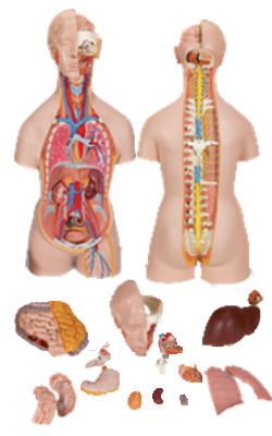
Main Model
TONGUE
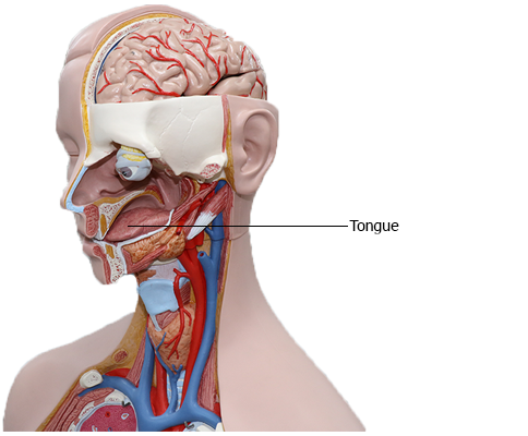
Tongue
The tongue (Latin lingua; Greek glossa) is a mobile muscular organ
covered with mucous membrane. It can assume a variety of
shapes and positions. It is partly in the oral cavity and partly in
the oropharynx. The tongue's main functions are articulation
(forming words during speaking) and squeezing food into the oropharynx as part of deglutition (swallowing). The tongue is
also involved with mastication, taste, and oral cleansing.
Parts and Surfaces of Tongue
The tongue has a root, body, and apex. The root
of the tongue is the attached posterior portion, extending between the mandible, hyoid, and the nearly vertical posterior surface of the tongue. The body of the tongue is
the anterior, approximately two thirds of the tongue between root and apex. The apex (tip) of the tongue is the anterior
end of the body, which rests against the incisor teeth. The
body and apex of the tongue are extremely mobile.
The tongue features two surfaces. The more extensive,
superior and posterior surface is the dorsum of the tongue
(commonly referred to as the "top" of the tongue). The inferior
surface of the tongue (commonly referred to as its "underside")
usually rests against the floor of the mouth. The margin of the
tongue separating the two surfaces is related on each side to the lingual gingivae and lateral teeth. The dorsum of the tongue
is characterized by a V-shaped groove, the terminal sulcus of
the tongue, the angle of which points posteriorly to the foramen cecum. This small pit, frequently absent, is
the non-functional remnant of the proximal part of the embryonic thyroglossal duct from which the thyroid gland developed.
The terminal sulcus divides the dorsum of the tongue transversely into a presulcal anterior part in the oral cavity proper
and a postsulcal posterior part in the oropharynx.
A midline groove divides the anterior part of the tongue
into right and left parts. The mucosa of the anterior part
of the tongue is relatively thin and closely attached to the
underlying muscle. It has a rough texture because of numerous small lingual papillae:
• Vallate papillae: large and flat topped, lie directly anterior to the terminal sulcus and are arranged in a V-shaped
row. They are surrounded by deep circular trenches, the
walls of which are studded with taste buds. The ducts of
the serous glands of the tongue open into the trenches.
• Foliate papillae: small lateral folds of the lingual mucosa.
They are poorly developed in humans.
• Filiform papillae: long and numerous, contain afferent
nerve endings that are sensitive to touch. These scaly, conical projections are pinkish gray and are arranged in V-shaped
rows that are parallel to the terminal sulcus, except at the
apex, where they tend to be arranged transversely.
• Fungiform papillae: mushroom shaped pink or red spots
scattered among the filiform papillae but most numerous
at the apex and margins of the tongue.
The vallate, foliate, and most of the fungiform papillae contain taste receptors in the taste buds.
The mucosa of the posterior part of the tongue is thick and
freely movable. It has no lingual papillae, but the underlying
lymphoid nodules give this part of the tongue an irregular, cobblestone appearance. The lymphoid nodules are known
collectively as the lingual tonsil. The pharyngeal part of the
tongue constitutes the anterior wall of the oropharynx and
can be inspected only with a mirror or downward pressure
on the tongue with a tongue depressor.
The inferior surface of the tongue is covered with a
thin, transparent mucous membrane. This surface is connected to the floor of the mouth by a midline fold
called the frenulum of the tongue. The frenulum allows
the anterior part of the tongue to move freely. On each side
of the frenulum, a deep lingual vein is visible through the
thin mucous membrane. A sublingual caruncle (papilla)
is present on each side of the base of the lingual frenulum
that includes the opening of the submandibular duct from
the submandibular salivary gland.
Muscles of Tongue
The tongue is essentially a mass of muscles that is mostly covered by mucosa (mucous membrane). As in the orbit, it is traditional to provide descriptions of the
actions of tongue muscles ascribing (1) a single action to a specific muscle, or (2) implying that a particular movement is the
consequence of a single muscle acting. This approach facilitates
learning, but greatly oversimplifies the actions of the tongue.
The muscles of the tongue do not act in isolation, and some
muscles perform multiple actions. Parts of a single muscle are
capable of acting independently, producing different, even
antagonistic actions. In general, extrinsic muscles alter the
position of the tongue, and intrinsic muscles alter its shape.
The four intrinsic and four extrinsic muscles in each half of
the tongue are separated by a median fibrous lingual septum,
which merges posteriorly with the lingual aponeurosis.
Extrinsic Muscles of Tongue
The extrinsic muscles of
the tongue (genioglossus, hyoglossus, styloglossus, and palatoglossus) originate outside the tongue and attach to it. They
mainly move the tongue but they can alter its shape as well.
Intrinsic Muscles of Tongue
The superior and inferior
longitudinal, transverse, and vertical muscles are confined to
the tongue. They have their attachments entirely within the
tongue and are not attached to bone. The superior and
inferior longitudinal muscles act together to make the
tongue short and thick and to retract the protruded tongue.
The transverse and vertical muscles act simultaneously
to make the tongue long and narrow, which may push the
tongue against the incisor teeth or protrude the tongue from
the open mouth (especially when acting with the posterior
inferior part of the genioglossus).
Innervation of Tongue
All muscles of the tongue, except the palatoglossus, receive
motor innervation from CN XII, the hypoglossal nerve. Palatoglossus is a palatine muscle supplied by the
pharyngeal plexus. For general sensation (touch and temperature), the mucosa of the anterior two
thirds of the tongue is supplied by the lingual nerve, a branch
of CN V3. For special sensation
(taste), this part of the tongue, except for the vallate papillae,
is supplied the chorda tympani nerve, a branch of CN VII. The
chorda tympani joins the lingual nerve in the infratemporal
fossa and runs anteriorly in its sheath. The mucosa of the posterior third of the tongue and the vallate papillae are supplied
by the lingual branch of the glossopharyngeal nerve (CN IX)
for both general and special sensation. Twigs of the
internal laryngeal nerve, a branch of the vagus nerve (CN
X), supply mostly general but some special sensation to a small
area of the tongue just anterior to the epiglottis. These mostly
sensory nerves also carry parasympathetic secretomotor
fibers to serous glands in the tongue.
There are four basic taste sensations: sweet, salty, sour,
and bitter. Sweetness is detected at the apex, saltiness at the
lateral margins, and sourness and bitterness at the posterior
part of the tongue. All other "tastes" expressed by gourmets
are olfactory (smell and aroma).
Vasculature of Tongue
The arteries of the tongue are derived from the lingual artery,
which arises from the external carotid artery. On
entering the tongue, the lingual artery passes deep to the hyoglossus muscle. The dorsal lingual arteries supply the root of
the tongue; the deep lingual arteries supply the lingual body.
The deep lingual arteries communicate with each other near
the apex of the tongue. The dorsal lingual arteries are prevented
from communicating by the lingual septum.
The veins of the tongue are the dorsal lingual veins,
which accompany the lingual artery; the deep lingual veins,
which begin at the apex of the tongue, run posteriorly beside
the lingual frenulum to join the sublingual vein. The sublingual veins in elderly people are often varicose (enlarged and tortuous). Some or all of them may drain into the
IJV, or they may do so indirectly, joining first to form a lingual
vein that accompanies the initial part of the lingual artery.
The lymphatic drainage of the tongue is exceptional.
Most of the lymphatic drainage converges toward and follows the venous drainage; however, lymph from the tip of the
tongue, frenulum, and central lower lip runs an independent
course. Lymph from the tongue takes four routes:
1. Lymph from the root drains bilaterally into the superior
deep cervical lymph nodes.
2. Lymph from the medial part of the body drains bilaterally
and directly to the inferior deep cervical lymph nodes.
3. Lymph from the right and left lateral parts of body drains to
the submandibular lymph nodes on the ipsilateral side.
4. The apex and frenulum drain to the submental lymph
nodes, the medial portion draining bilaterally.
All lymph from the tongue ultimately drains to the deep cervical nodes, and passes via the jugular venous trunks into the
venous system at the right and left venous angles.