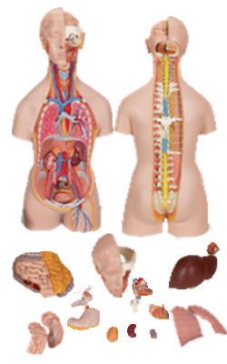
Main Model
SUBMANDIBULAR GLAND
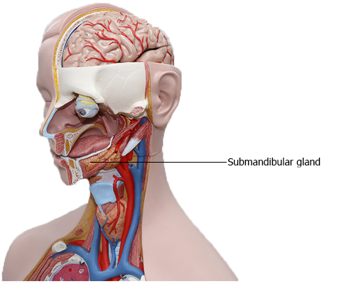
Salivary Glands
The salivary glands are the parotid, submandibular, and
sublingual glands. The clear, tasteless, odorless viscid fluid, saliva, secreted by these glands and the mucous
glands of the oral cavity:
• Keeps the mucous membrane of the mouth moist.
• Lubricates the food during mastication.
• Begins the digestion of starches.
• Serves as an intrinsic "mouthwash."
• Plays significant roles in the prevention of tooth decay and
in the ability to taste.
In addition to the main salivary glands, small accessory salivary glands are scattered over the palate, lips, cheeks, tonsils, and tongue. The parotid glands are the largest of the three
paired salivary glands. The parotid glands are located lateral and posterior
to the rami of the mandible and masseter muscles, within
unyielding fibrous sheaths. The parotid glands drain anteriorly via single ducts that enter the oral vestibule opposite the
second maxillary molar teeth.
Submandibular Glands
The submandibular glands lie along the body of the mandible, partly superior and partly inferior to the posterior half
of the mandible, and partly superficial and partly deep to the mylohyoid muscle. The submandibular duct,
approximately 5 cm long, arises from the portion of the gland
that lies between the mylohyoid and hyoglossus muscles.
Passing from lateral to medial, the lingual nerve loops under
the duct that runs anteriorly, opening by one to three orifices
on a small sublingual papilla beside the base of the lingual
frenulum. The orifices of the submandibular ducts are visible, and saliva can often be seen trickling from
them (or spraying from them during yawning). The arterial
supply of the submandibular glands is from the submental arteries. The veins accompany the arteries.
The lymphatic vessels of the glands end in the deep cervical lymph nodes, particularly the jugulo-omohyoid node.
The submandibular glands are supplied by presynaptic
parasympathetic secretomotor fibers conveyed from the
facial nerve to the lingual nerve by the chorda tympani nerve,
which synapse with postsynaptic neurons in the submandibular ganglion. The latter fibers accompany arteries
to reach the gland, along with vasoconstrictive postsynaptic
sympathetic fibers from the superior cervical ganglion.
Sublingual Glands
The sublingual glands are the smallest and most deeply situated of the salivary glands. Each almond-shaped
gland lies in the floor of the mouth between the mandible and
the genioglossus muscle. The glands from each side unite to
form a horseshoe-shaped mass around the connective tissue
core of the lingual frenulum. Numerous small sublingual
ducts open into the floor of the mouth along the sublingual
folds. The arterial supply of the sublingual glands is from
the sublingual and submental arteries, branches of the lingual and facial arteries, respectively. The nerves
of the glands accompany those of the submandibular gland. Presynaptic parasympathetic secretomotor fibers are conveyed by the facial, chorda tympani, and lingual nerves to
synapse in the submandibular ganglion.