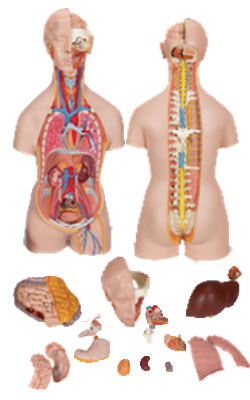
Main Model

HEART : Right pulmonary veins

Left Atrium
The left atrium forms most of the base of the heart. The valveless pairs of right and left pulmonary veins enter the smooth-walled atrium. In the embryo, there is only one common pulmonary vein, just as there is a single pulmonary trunk. The wall of this vein and four of its tributaries were incorporated into the wall of the left atrium, in the same way that the sinus venosus was incorporated into the right atrium. The part of the wall derived from the embryonic pulmonary vein is smooth walled. The tubular, muscular left auricle, its wall trabeculated with pectinate muscles, forms the superior part of the left border of the heart and overlaps the root of the pulmonary trunk. It represents the remains of the left part of the primordial atrium. A semilunar depression in the interatrial septum indicates the floor of the oval fossa; the surrounding ridge is the valve of the oval fossa (Latin valvulae foramen ovale).
The interior of the left atrium has
• A larger smooth-walled part and a smaller muscular auricle containing pectinate muscles.
• Four pulmonary veins (two superior and two inferior) entering its smooth posterior wall.
• A slightly thicker wall than that of the right atrium.
• An interatrial septum that slopes posteriorly and to the right.
• A left AV orifice through which the left atrium discharges the oxygenated blood it receives from the pulmonary veins into the left ventricle.