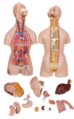
Main Model
KIDNEY : Right kidney (1)

Kidneys, Ureters, and Suprarenal Glands
The kidneys produce urine that is conveyed by the ureters to
the urinary bladder in the pelvis. The superomedial aspect
of each kidney normally contacts a suprarenal gland. A weak fascial septum separates the glands from the kidneys; thus
they are not actually attached to each other. The
suprarenal glands function as part of the endocrine system,
completely separate in function from the kidneys. The superior urinary organs (kidneys and ureters), their vessels, and
the suprarenal glands are primary retroperitoneal structures on the posterior abdominal wall - that is, they
were originally formed as and remain retroperitoneal viscera.
Perinephric fat (perirenal fat capsule) surrounds the kidneys and their vessels as it extends into their hollow centers, the
renal sinuses. The kidneys, suprarenal glands, and
the fat surrounding them are enclosed (except inferiorly) by a
condensed, membranous layer of renal fascia, which continues medially to ensheath the renal vessels, blending with the
vascular sheaths of the latter. Inferomedially, a delicate extension of the renal fascia is prolonged along the ureter as the peri-ureteric fascia. External to the renal fascia is paranephric
fat (pararenal fat body), the extraperitoneal fat of the lumbar
region that is most obvious posterior to the kidney. The renal fascia sends collagen bundles through the paranephric fat.
The collagen bundles, renal fascia, and perinephric and
paranephric fat, along with the tethering provided by the
renal vessels and ureter, hold the kidneys in a relatively
fixed position. However, movement of the kidneys occurs
during respiration and when changing from the supine to
the erect position, and vice versa. Normal renal mobility is approximately 3 cm, approximately the height of one vertebral
body. Superiorly, the renal fascia is continuous with the fascia
on the inferior surface of the diaphragm (diaphragmatic fascia);
thus the primary attachment of the suprarenal glands is to the
diaphragm. Inferiorly, the anterior and posterior layers of renal
fascia are only loosely united, if attached at all.
Kidneys
The ovoid kidneys remove excess water, salts, and wastes of
protein metabolism from the blood while returning nutrients
and chemicals to the blood. They lie retroperitoneally on the posterior abdominal wall, one on each side of the vertebral
column at the level of the T12-L3 vertebrae.
At the concave medial margin of each kidney is a vertical
cleft, the renal hilum. The renal hilum is the entrance to a space within the kidney, the renal sinus.
Structures that serve the kidneys (vessels, nerves, and structures
that drain urine from the kidney) enter and exit the renal sinus through the renal hilum. The hilum of the left kidney lies near
the transpyloric plane, approximately 5 cm from the median
plane. The transpyloric plane passes through the superior pole of the right kidney, which is approximately 2.5 cm
lower than the left pole, probably due to the presence of the
liver. Posteriorly, the superior parts of the kidneys lie deep to
the 11th and 12th ribs. The levels of the kidneys change during respiration and with changes in posture. Each kidney moves
2-3 cm in a vertical direction during the movement of the diaphragm that occurs with deep breathing. Because the usual surgical approach to the kidneys is through the posterior abdominal wall, it is helpful to know that the inferior pole of the right kidney is approximately a finger's breadth superior to the iliac crest.
During life, the kidneys are reddish brown and measure
approximately 10 cm in length, 5 cm in width, and 2.5 cm in
thickness. Superiorly, the posterior aspects of the kidneys are
associated with the diaphragm, which separates them from
the pleural cavities and the 12th pair of ribs. More
inferiorly, the posterior surfaces of the kidney are related to
the psoas major muscles medially and the quadratus lumborum muscle. The subcostal nerve and vessels and
the iliohypogastric and ilio-inguinal nerves descend diagonally
across the posterior surfaces of the kidneys. The liver, duodenum, and ascending colon are anterior to the right kidney. This kidney is separated from the liver
by the hepatorenal recess. The left kidney is related to the
stomach, spleen, pancreas, jejunum, and descending colon.
At the hilum, the renal vein is anterior to the renal artery,
which is anterior to the renal pelvis. Within the kidney, the renal sinus is occupied by the renal
pelvis, calices, vessels, and nerves, and a variable amount of
fat. Each kidney has anterior and posterior
surfaces, medial and lateral margins, and superior and inferior poles. However, because of the protrusion of the lumbar
vertebral column into the abdominal cavity, the kidneys are
obliquely placed, lying at an angle to each other.
Consequently, the transverse diameter of the kidneys is foreshortened in anterior views and anteroposterior
(AP) radiographs. The lateral margin of each kidney is convex, and the medial margin is concave where the
renal sinus and renal pelvis are located. The indented medial
margin gives the kidney a somewhat bean-shaped appearance.
The renal pelvis is the flattened, funnel-shaped expansion of the superior end of the ureter. The apex of the renal pelvis is continuous with
the ureter. The renal pelvis receives two or three major calices (calyces), each of which divides into two or three minor
calices. Each minor calyx is indented by a renal papilla, the
apex of the renal pyramid, from which the urine is excreted. In living persons, the renal pelvis and its calices are usually
collapsed (empty). The pyramids and their associated cortex
form the lobes of the kidney. The lobes are visible on the external surfaces of the kidneys in fetuses, and evidence of
the lobes may persist for some time after birth.
Vessels and Nerves of Kidneys
Renal Arteries and Veins
The renal arteries arise at the
level of the IV disc between the L1 and L2 vertebrae. The longer right renal artery passes posterior
to the IVC. Typically, each artery divides close to the hilum into
five segmental arteries that are end arteries (i.e., they do not
anastomose significantly with other segmental arteries, so that
the area supplied by each segmental artery is an independent,
surgically resectable unit or renal segment). Segmental arteries are distributed to the renal segments as follows:
• The superior (apical) segment is supplied by the superior (apical) segmental artery; the anterosuperior and
antero-inferior segments are supplied by the anterosuperior segmental and antero-inferior segmental arteries; and the inferior segment is supplied by the inferior segmental artery. These arteries originate from the
anterior branch of the renal artery.
• The posterior segmental artery, which originates from
a continuation of the posterior branch of the renal artery,
supplies the posterior segment of the kidney.
Multiple renal arteries are common and usually enter the
hilum of the kidney. Extrahilar renal arteries from
the renal artery or aorta may enter the external surface of the
kidney, commonly at their poles ("polar arteries").
Several renal veins drain each kidney and unite in a variable
fashion to form the right and left renal veins; these veins lie
anterior to the right and left renal arteries. The longer left renal vein receives the left suprarenal vein, the left gonadal (testicular
or ovarian) vein, and a communication with the ascending lumbar
vein; it then traverses the acute angle between the SMA anteriorly
and the aorta posteriorly. Each renal vein drains into the IVC.
Lymphatics of Kidneys
The renal lymphatic vessels follow the renal veins
and drain into the right and left lumbar (caval and aortic)
lymph nodes.
Nerves of Kidneys
The nerves to the kidneys arise from the renal
nerve plexus and consist of sympathetic and parasympathetic fibers. The renal nerve plexus is supplied by fibers from the abdominopelvic (especially the least)
splanchnic nerves.