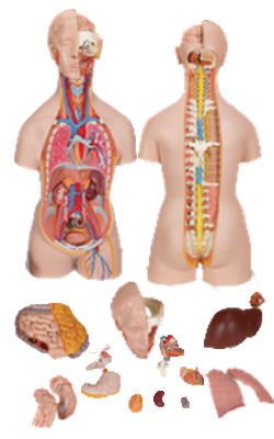
Main Model
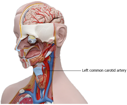
LEFT COMMON CAROTID ARTERY
Superior Mediastinum and Great Vessels
The superior mediastinum is superior to the transverse
thoracic plane, passing through the sternal angle and the
junction (IV disc) of vertebrae T4 and T5. From
anterior to posterior, the contents of the superior mediastinum are the:
• Thymus.
• Great vessels, with the veins (brachiocephalic veins and
SVC) anterior to the arteries (arch of aorta and roots of its
major branches - the brachiocephalic trunk, left common
carotid artery, and left subclavian artery) and related nerves
(vagus and phrenic nerves and the cardiac plexus of nerves).
• Inferior continuation of the cervical viscera (trachea anteriorly and esophagus posteriorly) and related nerves (left
recurrent laryngeal nerve).
• Thoracic duct and lymphatic trunks.
To summarize systemically, the order of the major structures
in the superior mediastinum, from anterior to posterior, is:
(1) thymus, (2) veins, (3) arteries, (4) airway, (5) alimentary
tract, and (6) lymphatic trunks.
Great Vessels
The right and left brachiocephalic veins are formed posterior to the sternoclavicular (SC) joints by the union of the
internal jugular and subclavian veins. At the level of the inferior border of the 1st right costal cartilage, the brachiocephalic veins unite to form the SVC.
The left brachiocephalic vein is more than twice as long as the right brachiocephalic vein because it passes from the
left to the right side, anterior to the roots of the three major
branches of the arch of the aorta. The brachiocephalic veins shunt blood from the head, neck, and upper
limbs to the right atrium.
The superior vena cava (SVC) returns blood from all
structures superior to the diaphragm, except the lungs and
heart. It passes inferiorly and ends at the level of the 3rd costal cartilage, where it enters the right atrium of the heart.
The SVC lies in the right side of the superior mediastinum,
anterolateral to the trachea and posterolateral to the ascending aorta. The right phrenic nerve lies between the SVC and
the mediastinal pleura. The terminal half of the SVC is in the
middle mediastinum, where it lies beside the ascending aorta
and forms the posterior boundary of the transverse pericardial sinus.
The ascending aorta, approximately 2.5 cm in diameter,
begins at the aortic orifice. Its only branches are the coronary
arteries, arising from the aortic sinuses. The
ascending aorta is intrapericardial; for this
reason, and because it lies inferior to the transverse thoracic
plane, it is considered a content of the middle mediastinum
(part of inferior mediastinum).
The arch of the aorta (aortic arch), the curved continuation of the ascending aorta, begins posterior to the 2nd right sternocostal (SC) joint
at the level of the sternal angle. It arches superiorly, posteriorly and to the left, and then inferiorly. The arch ascends
anterior to the right pulmonary artery and the bifurcation of
the trachea, reaching its apex at the left side of the trachea and esophagus as it passes over the root of the left lung. The arch
descends posterior to the left root of the lung beside the T4 vertebra. The arch ends by becoming the thoracic (descending) aorta posterior to the 2nd left sternocostal joint.
The arch of the azygos vein occupies a position corresponding to the aorta on the right side of the trachea over
the root of the right lung, although the blood is flowing in the
opposite direction. The ligamentum arteriosum, the remnant of the fetal ductus arteriosus, passes from the root of the left pulmonary artery to the inferior surface
of the arch of the aorta. The usual branches of the arch are
the brachiocephalic trunk, left common carotid artery, and
left subclavian artery.
The brachiocephalic trunk, the first and largest branch
of the arch of the aorta, arises posterior to the manubrium,
where it is anterior to the trachea and posterior to the left
brachiocephalic vein.
The trunk ascends superolaterally to reach the right side of
the trachea and the right SC joint, where it divides into the
right common carotid and right subclavian arteries.
The left common carotid artery, the second branch
of the arch of the aorta, arises posterior to the manubrium,
slightly posterior and to the left of the brachiocephalic trunk.
It ascends anterior to the left subclavian artery and is at first
anterior to the trachea and then to its left. It enters the neck
by passing posterior to the left SC joint.
The left subclavian artery, the third branch of the arch
of the aorta, arises from the posterior part of the arch, just
posterior to the left common carotid artery. It ascends lateral
to the trachea and left common carotid artery through the
superior mediastinum; it has no branches in the mediastinum. As it leaves the thorax and enters the root of the neck,
it passes posterior to the left SC joint.