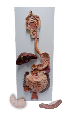
Main Model
Tooth
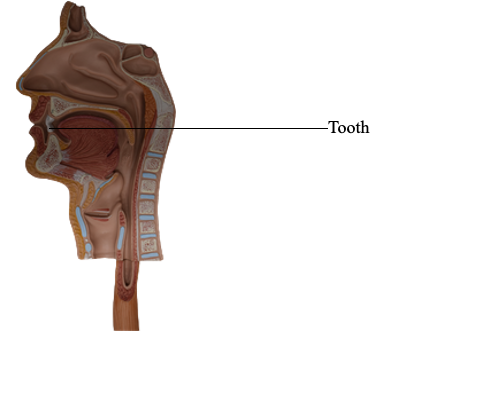
Teeth
The chief functions of teeth are to:
• Incise (cut), reduce, and mix food material with saliva
during mastication (chewing).
• Help sustain themselves in the tooth sockets by assisting
the development and protection of the tissues that support them.
• Participate in articulation (distinct connected speech).
The teeth are set in the tooth sockets and are used in mastication and in assisting in articulation. A tooth is identified and
described on the basis of whether it is deciduous (primary) or permanent (secondary), the type of tooth, and its proximity to the midline or front of the mouth (e.g., medial and
lateral incisors; the 1st molar is anterior to the 2nd).
Children have 20 deciduous teeth; adults normally have
32 permanent teeth. Before eruption, the
developing teeth reside in the alveolar arches as tooth buds.
The types of teeth are identified by their characteristics:
incisors, thin cutting edges; canines, single prominent
cones; premolars (bicuspids), two cusps; and molars, three
or more cusps. The vestibular surface
(labial or buccal) of each tooth is directed outwardly, and the
lingual surface is directed inwardly. As used in clinical (dental) practice, the mesial surface of a tooth is directed toward the median plane of the facial part of the cranium. The distal surface is directed away from this plane;
both mesial and distal surfaces are contact surfaces - that is, surfaces that contact adjacent teeth. The masticatory surface
is the occlusal surface.
Parts and Structure of Teeth
A tooth has a crown, neck, and root. The crown
projects from the gingiva. The neck is between the crown
and root. The root is fixed in the tooth socket by the periodontium (connective tissue surrounding roots); the number
of roots varies. Most of the tooth is composed of dentine (Latin dentinium), which is covered by enamel over the crown and cement (Latin cementum) over the root. The pulp cavity
contains connective tissue, blood vessels, and nerves. The
root canal (pulp canal) transmits the nerves and vessels to
and from the pulp cavity through the apical foramen.
The tooth sockets are in the alveolar processes of the
maxillae and mandible; they are the skeletal
features that display the greatest change during a lifetime. Adjacent sockets are separated by interalveolar septa; within the socket, the roots of teeth with more
than one root are separated by interradicular septa. The bone of the socket has a thin cortex
separated from the adjacent labial and lingual cortices by a variable amount of trabeculated bone. The labial wall of the
socket is particularly thin over the incisor teeth; the reverse
is true for the molars, where the lingual wall is thinner. Thus
the labial surface commonly is broken to extract incisors and
the lingual surface is broken to extract molars.
The roots of the teeth are connected to the bone of the alveolus by a springy suspension forming a special type of fibrous
joint called a dento-alveolar syndesmosis or gomphosis.
The periodontium (periodontal membrane) is composed of
collagenous fibers that extend between the cement of the root
and the periosteum of the alveolus. It is abundantly supplied
with tactile, pressoreceptive nerve endings, lymph capillaries,
and glomerular blood vessels that act as hydraulic cushioning
to curb axial masticatory pressure. Pressoreceptive nerve endings are capable of receiving changes in pressure as stimuli.
Vasculature of Teeth
The superior and inferior alveolar arteries, branches of the
maxillary artery, supply the maxillary and mandibular teeth,
respectively. The alveolar
veins have the same names and distribution accompany the
arteries. Lymphatic vessels from the teeth and gingivae pass
mainly to the submandibular lymph nodes.
Innervation of Teeth
The named branches of the superior (CN V2) and
inferior (CN V3) alveolar nerves give rise to dental plexuses
that supply the maxillary and mandibular teeth.