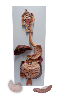
Main Model
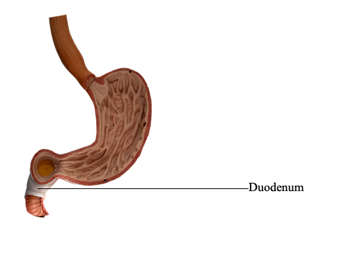
Duodenum
The small intestine, consisting of the duodenum, jejunum, and ileum, is the primary site for absorption of nutrients from ingested materials. It extends from the pylorus to the ileocecal junction where the ileum joins the cecum (the first part of the large intestine). The pyloric part of the stomach empties into the duodenum, duodenal admission being regulated by the pylorus.
The duodenum (Latin breadth of 12 fingers), the first and shortest (25 cm) part of the small intestine, is also the widest and most fixed part. The duodenum pursues a C-shaped course around the head of the pancreas. It begins at the pylorus on the right side and ends at the duodenojejunal flexure (junction) on the left side. This junction occurs approximately at the level of the L2 vertebra, 2-3 cm to the left of the midline. The junction usually takes the form of an acute angle, the duodenojejunal flexure. Most of the duodenum is fixed by peritoneum to structures on the posterior abdominal wall and is considered partially retroperitoneal.
The duodenum is divisible into four parts:
• Superior (first) part: short (approximately 5 cm) and lies anterolateral to the body of the L1 vertebra.
• Descending (second) part: longer (7-10 cm) and descends along the right sides of the L1-L vertebrae.
• Inferior (third) part: 6-8 cm long and crosses the L3 vertebra.
• Ascending (fourth) part: short (5 cm) and begins at the left of the L3 vertebra and rises superiorly as far as the superior border of the L2 vertebra.
The first 2 cm of the superior part of the duodenum, immediately distal to the pylorus, has a mesentery and is mobile. This free part, called the ampulla (duodenal cap), has an appearance distinct from the remainder of the duodenum when observed radiographically using contrast medium. The distal 3 cm of the superior part and the other three parts of the duodenum have no mesentery and are immobile because they are retroperitoneal.
The superior part of the duodenum ascends from the pylorus and is overlapped by the liver and gallbladder. Peritoneum covers its anterior aspect, but it is bare of peritoneum posteriorly, except for the ampulla. The proximal part has the hepatoduodenal ligament (part of the lesser omentum) attached superiorly and the greater omentum attached inferiorly.
The descending part of the duodenum runs inferiorly, curving around the head of the pancreas. Initially, it lies to the right of and parallel to the inferior vena cava (IVC). The bile and main pancreatic ducts enter its posteromedial wall. These ducts usually unite to form the hepatopancreatic ampulla, which opens on an eminence, called the major duodenal papilla, located posteromedially in the descending duodenum. The descending part of the duodenum is entirely retroperitoneal. The anterior surface of its proximal and distal thirds is covered with peritoneum; however, the peritoneum reflects from its middle third to form the double-layered mesentery of the transverse colon, the transverse mesocolon.
The inferior (horizontal) part of the duodenum runs transversely to the left, passing over the inferior vena cava (IVC), aorta, and L3 vertebra. It is crossed by the superior mesenteric artery and vein and the root of the mesentery of the jejunum and ileum. Superior to it is the head of the pancreas and its uncinate process. The anterior surface of the inferior part is covered with peritoneum, except where it is crossed by the superior mesenteric vessels and the root of the mesentery. Posteriorly it is separated from the vertebral column by the right psoas major, inferior vena cava (IVC), aorta, and the right testicular or ovarian vessels.
The ascending part of the duodenum runs superiorly and along the left side of the aorta to reach the inferior border of the body of the pancreas. Here it curves anteriorly to join the jejunum at the duodenojejunal flexure, supported by the attachment of a suspensory muscle of the duodenum (ligament of Treitz). This muscle is composed of a slip of skeletal muscle from the diaphragm and a fibromuscular band of smooth muscle from the third and fourth parts of the duodenum. Contraction of this muscle widens the angle of the duodenojejunal flexure, facilitating movement of the intestinal contents. The suspensory muscle passes posterior to the pancreas and splenic vein and anterior to the left renal vein.
The arteries of the duodenum arise from the celiac trunk and the superior mesenteric artery. The celiac trunk, via the gastroduodenal artery and its branch, the superior pancreaticoduodenal artery, supplies the duodenum proximal to the entry of the bile duct into the descending part of the duodenum. The superior mesenteric artery, through its branch, the inferior pancreaticoduodenal artery, supplies the duodenum distal to the entry of the bile duct. The pancreaticoduodenal arteries lie in the curve between the duodenum and the head of the pancreas and supply both structures. The anastomosis of the superior and inferior pancreaticoduodenal arteries (i.e., between the celiac and superior mesenteric arteries) occurs between the entry of the bile duct and the junction of the descending and inferior parts of the duodenum. An important transition in the blood supply of the digestive tract occurs here: proximally, extending orad (toward the mouth) to and including the abdominal part of the esophagus, the blood is supplied to the digestive tract by the celiac trunk; distally, extending aborad (away from the mouth) to the left colic flexure, the blood is supplied by the superior mesenteric artery (SMA). The basis of this transition in blood supply is embryological; this is the junction of the foregut and midgut.
The veins of the duodenum follow the arteries and drain into the hepatic portal vein, some directly and others indirectly, through the superior mesenteric and splenic veins. The lymphatic vessels of the duodenum follow the arteries.
The anterior lymphatic vessels drain into the pancreaticoduodenal lymph nodes, located along the superior and inferior pancreaticoduodenal arteries, and into the pyloric lymph nodes, which lie along the gastroduodenal artery. The posterior lymphatic vessels pass posterior to the head of the pancreas and drain into the superior mesenteric lymph nodes. Efferent lymphatic vessels from the duodenal lymph nodes drain into the celiac lymph nodes.
The nerves of the duodenum derive from the vagus and greater and lesser (abdominopelvic) splanchnic nerves by way of the celiac and superior mesenteric plexuses. The nerves are next conveyed to the duodenum via peri-arterial plexuses extending to the pancreaticoduodenal arteries.