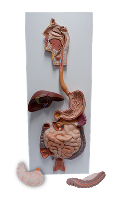
Main Model
Pancreas
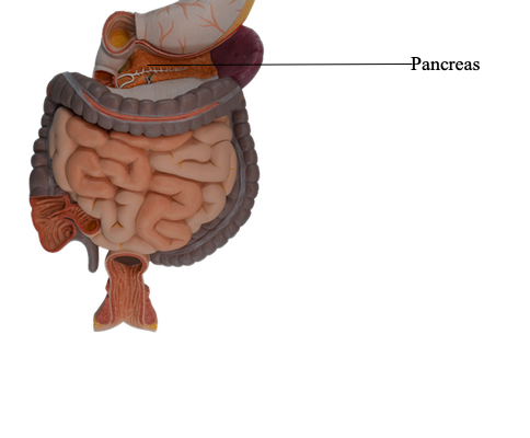
Pancreas
The pancreas is an elongated, accessory digestive gland that
lies retroperitoneally, overlying and transversely crossing the
bodies of the L1 and L2 vertebra (the level of the transpyloric plane) on the posterior abdominal wall. It
lies posterior to the stomach between the duodenum on the
right and the spleen on the left. The transverse
mesocolon attaches to its anterior margin.
The pancreas produces:
• an exocrine secretion (pancreatic juice from the acinar
cells) that enters the duodenum through the main and
accessory pancreatic ducts.
• endocrine secretions (glucagon and insulin from the
pancreatic islets [of Langerhans]) that enter the blood.
For descriptive purposes, the pancreas is divided into four
parts: head, neck, body, and tail.
The head of the pancreas is the expanded part of the
gland that is embraced by the C-shaped curve of the duodenum to the right of the superior mesenteric vessels just
inferior to the transpyloric plane. It firmly attaches to the
medial aspect of the descending and horizontal parts of
the duodenum. The uncinate process, a projection from
the inferior part of the pancreatic head, extends medially
to the left, posterior to the SMA. The pancreatic head rests posteriorly on the IVC, right renal artery
and vein, and left renal vein. On its way to opening into
the descending part of the duodenum, the bile duct lies
in a groove on the posterosuperior surface of the head or is embedded in its substance.
The neck of the pancreas is short (1.5-2 cm) and overlies the superior mesenteric vessels, which form a groove
in its posterior aspect. The anterior
surface of the neck, covered with peritoneum, is adjacent
to the pylorus of the stomach. The SMV joins the splenic
vein posterior to the neck to form the hepatic portal vein.
The body of the pancreas continues from the neck and lies
to the left of the superior mesenteric vessels, passing over the
aorta and L2 vertebra, continuing just above the transpyloric
plane posterior to the omental bursa. The anterior surface of
the body of the pancreas is covered with peritoneum and lies in the floor of the omental bursa and forms part of the stomach
bed. The posterior surface of the body is
devoid of peritoneum and is in contact with the aorta, SMA,
left suprarenal gland, left kidney, and renal vessels.
The tail of the pancreas lies anterior to the left kidney,
where it is closely related to the splenic hilum and the left colic
flexure. The tail is relatively mobile and passes between the layers
of the splenorenal ligament with the splenic vessels.
The main pancreatic duct begins in the tail of the pancreas and runs through the parenchyma of the gland to the
pancreatic head: here it turns inferiorly and is closely related
to the bile duct. The main pancreatic duct
and bile duct usually unite to form the short, dilated hepatopancreatic ampulla (of Vater), which opens into the
descending part of the duodenum at the summit of the major
duodenal papilla. At least 25% of the time,
the ducts open into the duodenum separately.
The sphincter of the pancreatic duct (around the
terminal part of the pancreatic duct), the sphincter of the
bile duct (around the termination of the bile duct), and the
hepatopancreatic sphincter (of Oddi) - around the hepatopancreatic ampulla - are smooth muscle sphincters that control the flow of bile and pancreatic juice into the ampulla
and prevent reflux of duodenal content into the ampulla.
The accessory pancreatic duct opens into
the duodenum at the summit of the minor duodenal papilla. Usually, the accessory duct communicates with
the main pancreatic duct. In some cases, the main pancreatic
duct is smaller than the accessory pancreatic duct and the
two may not be connected. In such cases, the accessory duct
carries most of the pancreatic juice.
The arterial supply of the pancreas is derived mainly from
the branches of the markedly tortuous splenic artery. Multiple pancreatic arteries form several arcades with pancreatic branches of the gastroduodenal and superior mesenteric
arteries. As many as 10 branches may pass from
the splenic artery to the body and tail of the pancreas. The
anterior and posterior superior pancreaticoduodenal arteries, branches of the gastroduodenal artery, and the anterior and posterior inferior pancreaticoduodenal arteries,
branches of the SMA, form anteriorly and posteriorly placed
arcades that supply the head of the pancreas.
Venous drainage from the pancreas occurs via corresponding pancreatic veins, tributaries of the splenic and superior
mesenteric parts of the hepatic portal vein; most empty into
the splenic vein.
The pancreatic lymphatic vessels follow the blood vessels. Most vessels end in the pancreaticosplenic
lymph nodes, which lie along the splenic artery. Some vessels
end in the pyloric lymph nodes. Efferent vessels from these
nodes drain to the superior mesenteric lymph nodes or to the
celiac lymph nodes via the hepatic lymph nodes.
The nerves of the pancreas are derived from the vagus and
abdominopelvic splanchnic nerves passing through the diaphragm. The parasympathetic and sympathetic
fibers reach the pancreas by passing along the arteries from
the celiac plexus and superior mesenteric plexus. In addition to sympathetic fibers that pass to blood vessels,
sympathetic and parasympathetic fibers are distributed to pancreatic acinar cells and islets. The parasympathetic fibers
are secretomotor, but pancreatic secretion is primarily mediated by secretin and cholecystokinin, hormones formed by
the epithelial cells of the duodenum and proximal intestinal
mucosa under the stimulus of acid contents from the stomach.