Main Model
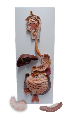
Spleen
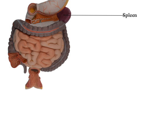
Spleen
The spleen is an ovoid, usually purplish, pulpy mass about
the size and shape of one's fist. It is relatively delicate and
considered the most vulnerable abdominal organ. The spleen
is located in the superolateral part of the left upper quadrant
(LUQ), or hypochondrium of the abdomen, where it enjoys
protection of the inferior thoracic cage.
As the largest of the lymphatic organs, it participates in the
body's defense system as a site of lymphocyte (white blood
cell) proliferation and of immune surveillance and response.
Prenatally, the spleen is a hematopoietic (blood-forming)
organ, but after birth it is involved primarily in identifying,
removing, and destroying expended red blood cells (RBCs)
and broken-down platelets, and in recycling iron and globin.
The spleen serves as a blood reservoir, storing RBCs and platelets, and, to a limited degree, can provide a sort of "self-transfusion" as a response to the stress imposed by hemorrhage. In
spite of its size and the many useful and important functions
it provides, it is not a vital organ (not necessary to sustain life).
To accommodate these functions, the spleen is a soft, vascular (sinusoidal) mass with a relatively delicate fibroelastic
capsule. The thin capsule is covered with a layer
of visceral peritoneum that entirely surrounds the spleen
except at the splenic hilum, where the splenic branches
of the splenic artery and vein enter and leave.
Consequently, it is capable of marked expansion and some
relatively rapid contraction.
The spleen is a mobile organ although it normally does
not descend inferior to the costal (rib) region; it rests on the
left colic flexure. It is associated posteriorly
with the left 9th-11th ribs (its long axis is roughly parallel
to the 10th rib) and separated from them by the diaphragm
and the costodiaphragmatic recess - the cleft-like extension of the pleural cavity between the diaphragm and the lower
part of the thoracic cage. The relations of the spleen are:
• Anteriorly, the stomach
• Posteriorly, the left part of the diaphragm, which separates it from the pleura, lung, and ribs 9-11
• Inferiorly, the left colic flexure
• Medially, the left kidney.
The spleen varies considerably in size, weight, and shape; however, it is usually approximately 12 cm long and 7 cm wide. (A
nonmetric memory device exploits odd numbers: the spleen is
1 inch thick, 3 inches wide, 5 inches long, and weighs 7 ounces.)
The diaphragmatic surface of the spleen is convexly
curved to fit the concavity of the diaphragm and curved bodies of the adjacent ribs. The close relationship of the spleen to the ribs that normally protect it can
be a detrimental one in the presence of rib fractures. The anterior
and superior borders of the spleen are sharp and often
notched, whereas its posterior (medial) end and inferior
border are rounded. Normally, the spleen does
not extend inferior to the left costal margin; thus it is seldom
palpable through the anterolateral abdominal wall unless it
is enlarged. When it is hardened and enlarged to approximately three times its normal size, it moves inferior to the
left costal margin, and its superior (notched) border lies inferomedially. The notched border is helpful when palpating an enlarged spleen because when the person takes a deep
breath, the notches can often be palpated.
The spleen normally contains a large quantity of blood
that is expelled periodically into the circulation by the action
of the smooth muscle in its capsule and trabeculae. The large
size of the splenic artery (or vein) indicates the volume of blood that passes through the spleen's capillaries and sinuses.
The thin fibrous capsule of the spleen is composed of
dense, irregular, fibroelastic connective tissue that is thickened at the splenic hilum. Internally the trabeculae (small fibrous bands), arising from the deep aspect of
the capsule, carry blood vessels to and from the parenchyma
or splenic pulp, the substance of the spleen.
The spleen contacts the posterior wall of the stomach and
is connected to its greater curvature by the gastrosplenic ligament, and to the left kidney by the splenorenal ligament.
These ligaments, containing splenic vessels, are attached to
the hilum of the spleen on its medial aspect.
The splenic hilum is often in contact with the tail of the pancreas and constitutes the left boundary of the omental bursa.
The arterial supply of the spleen is from the splenic
artery, the largest branch of the celiac trunk.
It follows a tortuous course posterior to the omental bursa, anterior to the left kidney, and along the superior border of
the pancreas. Between the layers of the splenorenal ligament, the splenic artery divides into five or more branches
that enter the hilum. The lack of anastomosis of these arterial
vessels within the spleen results in the formation of vascular segments of the spleen: two in 84% of spleens and three in
the others, with relatively avascular planes between them,
enabling subtotal splenectomy.
Venous drainage from the spleen flows via the splenic vein,
formed by several tributaries that emerge from the hilum. It is joined by the IMV and runs posterior to the body and tail of the pancreas throughout most of
its course. The splenic vein unites with the SMV posterior to
the neck of the pancreas to form the hepatic portal vein.
The splenic lymphatic vessels leave the lymph nodes in
the splenic hilum and pass along the splenic vessels to the pancreaticosplenic lymph nodes en route to the celiac
nodes. The pancreaticosplenic nodes relate to
the posterior surface and superior border of the pancreas.
The nerves of the spleen, derived from the celiac plexus, are distributed mainly along branches of the
splenic artery, and are vasomotor in function.