Main Model
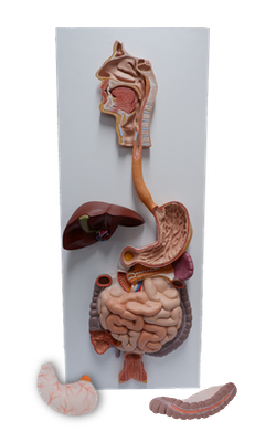
Pharynx
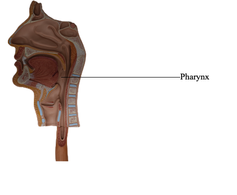
Pharynx
The pharynx is the superior expanded part of the alimentary system posterior to the nasal and oral cavities, extending
inferiorly past the larynx. The pharynx
extends from the cranial base to the inferior border of the cricoid cartilage anteriorly and the inferior border of the C6 vertebra posteriorly. The pharynx is widest (approximately
5 cm) opposite the hyoid and narrowest (approximately
1.5 cm) at its inferior end, where it is continuous with the
esophagus. The flat posterior wall of the pharynx lies against
the prevertebral layer of deep cervical fascia.
Interior of Pharynx
The pharynx is divided into three
parts:
• Nasopharynx: posterior to the nose and superior to the
soft palate.
• Oropharynx: posterior to the mouth.
• Laryngopharynx: posterior to the larynx.
The nasopharynx has a respiratory function; it is the posterior extension of the nasal cavities. The nose
opens into the nasopharynx through two choanae (paired
openings between the nasal cavity and the nasopharynx). The
roof and posterior wall of the nasopharynx form a continuous surface that lies inferior to the body of the sphenoid bone and
the basilar part of the occipital bone.
The abundant lymphoid tissue in the pharynx forms an
incomplete tonsillar ring around the superior part of the pharynx. The lymphoid
tissue is aggregated in certain regions to form masses called
tonsils. The pharyngeal tonsil (commonly called the adenoid
when enlarged) is in the mucous membrane of the roof and posterior wall of the nasopharynx. Extending inferiorly from the medial end of the pharyngotympanic
tube is a vertical fold of mucous membrane, the salpingopharyngeal fold. It covers the salpingopharyngeus muscle, which opens the pharyngeal orifice of the
pharyngotympanic tube during swallowing. The collection of
lymphoid tissue in the submucosa of the pharynx near the
nasopharyngeal opening, or orifice of the pharyngotympanic
tube, is the tubal tonsils. Posterior to the torus
of the pharyngotympanic tube and the salpingopharyngeal
fold is a slit-like lateral projection of the pharynx, the pharyngeal recess, which extends laterally and posteriorly.
The oropharynx has a digestive function. It is bounded
by the soft palate superiorly, the base of the tongue inferiorly,
and the palatoglossal and palatopharyngeal arches laterally. It extends from the soft palate to the
superior border of the epiglottis.
Deglutition (swallowing) is the complex process that
transfers a food bolus from the mouth through the pharynx
and esophagus into the stomach. Solid food is masticated
(chewed) and mixed with saliva to form a soft bolus (mass)
that is easier to swallow. Deglutition occurs in three stages:
• Stage 1: voluntary; the bolus is compressed against the
palate and pushed from the mouth into the oropharynx,
mainly by movements of the muscles of the tongue and
soft palate.
• Stage 2: involuntary and rapid; the soft palate is elevated,
sealing off the nasopharynx from the oropharynx and
laryngopharynx. The pharynx widens and
shortens to receive the bolus of food as the suprahyoid
muscles and longitudinal pharyngeal muscles contract,
elevating the larynx.
• Stage 3: involuntary; sequential contraction of all three
pharyngeal constrictor muscles creates a peristaltic ridge
that forces the food bolus inferiorly into the esophagus.
The palatine tonsils are collections of lymphoid tissue on
each side of the oropharynx in the interval between the palatine arches. The tonsil does not fill
the tonsillar sinus (fossa) between the palatoglossal and
palatopharyngeal arches in adults. The submucosal tonsillar bed, in which the palatine tonsil lies, is between these
arches. The tonsillar bed is formed by the superior pharyngeal constrictor and the thin, fibrous sheet
of pharyngobasilar fascia. This fascia
blends with the periosteum of the cranial base and defines
the limits of the pharyngeal wall in its superior part.
The laryngopharynx lies posterior to the larynx, extending from the superior border of the epiglottis and the pharyngo-epiglottic folds to the inferior border of the cricoid cartilage, where it narrows and becomes
continuous with the esophagus. Posteriorly, the laryngopharynx is related to the bodies of the C4-C6 vertebrae. Its posterior and lateral walls are formed by the middle and inferior
pharyngeal constrictor muscles. Internally, the
wall is formed by the palatopharyngeus and stylopharyngeus
muscles. The laryngopharynx communicates with the larynx
through the laryngeal inlet on its anterior wall.
The piriform fossa (recess) is a small depression of the
laryngopharyngeal cavity on either side of the laryngeal inlet.
This mucosa-lined fossa is separated from the laryngeal inlet
by the ary-epiglottic fold. Laterally, the piriform fossa is
bounded by the medial surfaces of the thyroid cartilage and
the thyrohyoid membrane. Branches of the internal laryngeal and recurrent laryngeal nerves lie deep to the
mucous membrane of the piriform fossa and are vulnerable
to injury when a foreign body lodges in the fossa.
Pharyngeal Muscles
The wall of the pharynx is exceptional for the alimentary tract, having a muscular layer composed
entirely of voluntary muscle, arranged with longitudinal muscles internal to a circular layer of muscles. Most of the alimentary
tract is composed of smooth muscle, with a layer of longitudinal
muscle external to a circular layer. The external circular layer of
pharyngeal muscles consists of three pharyngeal constrictors:
superior, middle, and inferior.
The internal longitudinal muscles consists of the palatopharyngeus, stylopharyngeus, and salpingopharyngeus. These
muscles elevate the larynx and shorten the pharynx during swallowing and speaking.
The pharyngeal constrictors have a strong internal fascial
lining, the pharyngobasilar fascia, and a thin
external fascial lining, the buccopharyngeal fascia.
Inferiorly, the buccopharyngeal fascia blends with the pretracheal layer of the deep cervical fascia. The pharyngeal
constrictors contract involuntarily so that contraction takes
place sequentially from the superior to the inferior end of
the pharynx, propelling food into the esophagus. All three
pharyngeal constrictors are supplied by the pharyngeal plexus of nerves that is formed by pharyngeal branches of
the vagus and glossopharyngeal nerves and by sympathetic
branches from the superior cervical ganglion. The pharyngeal plexus lies on the lateral wall of
the pharynx, mainly on the middle pharyngeal constrictor.
The overlapping of the pharyngeal constrictor muscles
leaves four gaps in the musculature for structures to enter or
leave the pharynx:
1. Superior to the superior pharyngeal constrictor, the levator veli palatini, pharyngotympanic tube, and ascending
palatine artery pass through a gap between the superior
pharyngeal constrictor and the cranium. It is here that the
pharyngobasilar fascia blends with the buccopharyngeal
fascia to form, with the mucous membrane, the thin wall
of the pharyngeal recess.
2. A gap between the superior and middle pharyngeal constrictors forms a passageway that allows the stylopharyngeus, glossopharyngeal nerve, and stylohyoid ligament to
pass to the internal aspect of the pharyngeal wall.
3. A gap between the middle and inferior pharyngeal constrictors allows the internal laryngeal nerve and superior
laryngeal artery and vein to pass to the larynx.
4. A gap inferior to the inferior pharyngeal constrictor allows
the recurrent laryngeal nerve and inferior laryngeal artery
to pass superiorly into the larynx.
Vessels of Pharynx
A branch of the facial artery, the
tonsillar artery passes through the superior pharyngeal constrictor muscle and enters the inferior pole of the
palatine tonsil. The tonsil also receives arterial twigs from the
ascending palatine, lingual, descending palatine, and ascending pharyngeal arteries. The large external palatine vein (paratonsillar vein) descends from the soft palate and passes close to
the lateral surface of the tonsil before it enters the pharyngeal venous plexus.
The tonsillar lymphatic vessels pass laterally and
inferiorly to the lymph nodes near the angle of the mandible and the jugulodigastric node, referred to as the
tonsillar node because of its frequent enlargement when
the tonsil is inflamed (tonsillitis). The palatine,
lingual, and pharyngeal tonsils form the pharyngeal lymphatic (tonsillar) ring, an incomplete circular band of
lymphoid tissue around the superior part of the pharynx. The antero-inferior part of the ring is formed
by the lingual tonsil in the posterior part of the tongue.
Lateral parts of the ring are formed by the palatine and
tubal tonsils, and posterior and superior parts are formed
by the pharyngeal tonsil.
Pharyngeal Nerves
The nerve supply to the pharynx
(motor and most of sensory) derives from the pharyngeal plexus of nerves. Motor fibers in the plexus are
derived from the vagus nerve (CN X) via its pharyngeal branch
or branches. They supply all muscles of the pharynx and soft
palate, except the stylopharyngeus (supplied by CN IX) and the
tensor veli palatini (supplied by CN V3). The inferior pharyngeal
constrictor also receives some motor fibers from the external
and recurrent laryngeal branches of the vagus. Sensory fibers in the plexus are derived from the glossopharyngeal nerve. They
are distributed to all three parts of the pharynx. In addition,
the mucous membrane of the anterior and superior nasopharynx receives innervation from the maxillary nerve (CN V2). The
tonsillar nerves are derived from the tonsillar plexus of nerves formed by branches of the glossopharyngeal and vagus nerves.