Main Model
Inner layer of eyeball : 15 Lens

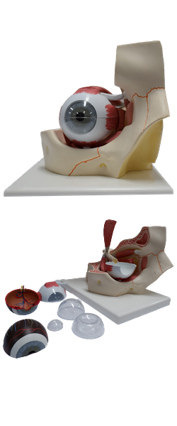
Refractive Media and Compartments of Eyeball
On their way to the retina, lightwaves pass through the
refractive media of the eyeball: cornea, aqueous humor, lens,
and vitreous humor. The cornea is the primary
refractory medium of the eyeball - that is, it bends light to
the greatest degree, focusing an inverted image on the light-sensitive retina of the fundus of the eyeball.
The aqueous humor (often shortened clinically to "aqueous") occupies the anterior segment of the eyeball. The anterior segment is subdivided
by the iris and pupil. The anterior chamber of the eye is
the space between the cornea anteriorly and the iris/pupil
posteriorly. The posterior chamber of the eye is between
the iris/pupil anteriorly and the lens and ciliary body posteriorly. Aqueous humor is produced in the posterior chamber
by the ciliary processes of the ciliary body. This clear watery
solution provides nutrients for the avascular cornea and lens.
After passing through the pupil into the anterior chamber,
the aqueous humor drains through a trabecular meshwork
at the iridocorneal angle into the scleral venous sinus
(Latin sinus venosus sclerae, canal of Schlemm). The
humor is removed by the limbal plexus, a network of scleral
veins close to the limbus, which drain in turn into both tributaries of the vorticose and anterior ciliary veins.
Intra-ocular pressure (IOP) is a balance between production
and outflow of aqueous humor.
The lens is posterior to the iris and anterior to the vitreous humor of the vitreous body. It is
a transparent, biconvex structure enclosed in a capsule. The
highly elastic capsule of the lens is anchored by zonular
fibers (collectively constituting the suspensory ligament
of the lens) to the encircling ciliary processes. Although
most refraction is produced by the cornea, the convexity of
the lens, particularly its anterior surface, constantly varies to
fine-tune the focus of near or distant objects on the retina. The isolated unattached lens assumes a nearly
spherical shape. In other words, in the absence of external attachment and stretching, it becomes nearly round. The ciliary muscle of the ciliary body changes the shape
of the lens. In the absence of nerve stimulation, the diameter of
the relaxed muscular ring is larger. The lens suspended within
the ring is under tension as its periphery is stretched, causing
it to be thinner (less convex). The less convex lens brings more
distant objects into focus (far vision). Parasympathetic stimulation via the oculomotor nerve (CN III) causes sphincter-like
contraction of the ciliary muscle. The ring becomes smaller,
and tension on the lens is reduced. The relaxed lens thickens (becomes more convex), bringing near objects into focus
(near vision). The active process of changing the shape of the
lens for near vision is called accommodation. The thickness
of the lens increases with aging so that the ability to accommodate typically becomes restricted after age 40.
The vitreous humor is a watery fluid enclosed in the
meshes of the vitreous body, a transparent jelly-like substance in the posterior four fifths of the eyeball posterior to
the lens (posterior segment of the eyeball, also called
the postremal or vitreous chamber). In addition
to transmitting light, the vitreous humor holds the retina in
place and supports the lens.