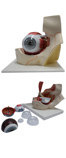
Main Model
Orbit : 30 Sphenoidal sinus

Paranasal Sinuses
The paranasal sinuses are air-filled extensions of the respiratory part of the nasal cavity into the following cranial bones: frontal, ethmoid, sphenoid, and maxilla. They are named according to the bones in which they are located. The sinuses continue to invade the surrounding bone, and marked extensions are common in the crania of older individuals.
Sphenoidal Sinuses
The sphenoidal sinuses are located in the body of the sphenoid, but they may extend into the wings of this bone. They are unevenly divided and separated by a bony septum. Because of this extensive pneumatization (formation of air cells), the body of the sphenoid is fragile. Only thin plates of bone separate the sinuses from several important structures: the optic nerves and optic chiasm, the pituitary gland, the internal carotid arteries, and the cavernous sinuses. The sphenoidal sinuses are derived from a posterior ethmoidal cell that begins to invade the sphenoid at approximately 2 years of age. In some people, several posterior ethmoidal cells invade the sphenoid, giving rise to multiple sphenoidal sinuses that open separately into the sphenoethmoidal recess. The posterior ethmoidal arteries and the posterior ethmoidal nerves that accompany the arteries supply the sphenoidal sinuses.