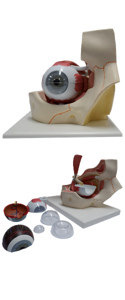
Main Model
Uvea : 7 Ciliary nerves

Nerves of Orbit
The large optic nerves convey purely sensory nerves that
transmit impulses generated by optical stimuli. They are cranial nerves (CN II) by convention,
but develop as paired anterior extensions of the forebrain
and are actually central nervous system (CNS) fiber tracts
formed of second-order neurons. The optic nerves begin
at the lamina cribrosa of the sclera, where the unmyelinated nerve fibers pierce the sclera and become myelinated, posterior to the optic disc. They exit the orbits via the
optic canals. Throughout their course in the orbit, the optic nerves are surrounded by extensions of the cranial meninges and subarachnoid space, the latter occupied by a thin
layer of CSF. The intra-orbital extensions of the cranial dura and arachnoid constitute the optic
nerve sheath, which becomes continuous anteriorly with
the fascial sheath of the eyeball and the sclera. A layer of
pia mater covers the surface of the optic nerve within the
sheath.
In addition to the optic nerve (CN II), the nerves of the
orbit include those that enter through the superior orbital
fissure and supply the ocular muscles: oculomotor (CN
III), trochlear (CN IV), and abducent (CN VI) nerves. A memory device for the innervation
of the extra-ocular muscles moving the eyeball is similar
to a chemical formula: LR6SO4AO3 (lateral rectus, CN VI; superior oblique, CN IV; all others, CN III). The trochlear and abducent nerves pass directly to the single muscle
supplied by each nerve. The oculomotor nerve divides into
a superior and an inferior division. The superior division
supplies the superior rectus and levator palpebrae superioris. The inferior division supplies the medial and inferior
rectus and inferior oblique and carries presynaptic parasympathetic fibers to the ciliary ganglion. The
movements are stimulated by the oculomotor, trochlear,
and abducent nerves, starting from the primary position in
the right and left orbits, and produce binocular vision.
The three terminal branches of the ophthalmic nerve, CN
V1 (the frontal, nasociliary, and lacrimal nerves), pass through
the superior orbital fissure and supply structures related to
the anterior orbit (e.g., lacrimal gland and eyelids), face, and
scalp.
The ciliary ganglion is a small group of postsynaptic
parasympathetic nerve cell bodies associated with CN V1.
It is located between the optic nerve and the lateral rectus
toward the posterior limit of the orbit. The ganglion receives
nerve fibers from three sources:
• Sensory fibers from CN V1 via the sensory or nasociliary
root of the ciliary ganglion.
• Presynaptic parasympathetic fibers from CN III via the
parasympathetic or oculomotor root of the ciliary
ganglion.
• Postsynaptic sympathetic fibers from the internal carotid
plexus via the sympathetic root of the ciliary ganglion.
The short ciliary nerves arise from the ciliary ganglion
and are considered to be branches of CN V1. They carry parasympathetic and sympathetic fibers to
the ciliary body and iris. The short ciliary nerves consist of
postsynaptic parasympathetic fibers originating in the ciliary ganglion, afferent fibers from the nasociliary nerve that pass
through the ganglion, and postsynaptic sympathetic fibers
that also pass through it. Long ciliary nerves, branches of the nasociliary nerve (CN V1) that pass to the eyeball, bypassing the ciliary ganglion, convey postsynaptic sympathetic
fibers to the dilator pupillae and afferent fibers from the iris
and cornea.
The posterior and anterior ethmoidal nerves, branches of
the nasociliary nerve arising in the orbit, exit via openings in
the medial wall of the orbit to supply the mucous membrane
of the sphenoidal and ethmoidal sinuses and the nasal cavities, as well as the dura of the anterior cranial fossa.