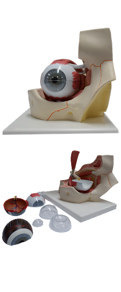
Main Model

Uvea : 8 Vorticose vein

Vasculature of Orbit
Arteries of Orbit
The blood supply of the orbit is mainly from the ophthalmic artery, a branch of the internal carotid artery
(Fig. 7.61; Table 7.9); the infra-orbital artery, from the external carotid artery, also contributes blood to structures
related to the orbital fl oor. The central artery of the retina, a branch of the ophthalmic artery arising inferior to
the optic nerve, pierces the sheath of the optic nerve and
runs within the nerve to the eyeball, emerging at the optic
disc (Fig. 7.45A, inset). Its branches spread over the internal surface of the retina (Figs. 7.52 and 7.62). The terminal
branches are end arteries (arterioles), which provide the only
blood supply to the internal aspect of the retina.
The external aspect of the retina is also supplied by the
capillary lamina of the choroid (choriocapillaris). Of the
eight or so posterior ciliary arteries (also branches of the ophthalmic artery), six short posterior ciliary arteries directly
supply the choroid, which nourishes the outer non-vascular
layer of the retina. Two long posterior ciliary arteries, one
on each side of the eyeball, pass between the sclera and the
choroid to anastomose with the anterior ciliary arteries
(continuations of the muscular branches of the ophthalmic artery to the rectus muscles) to supply the ciliary plexus.
Veins of Orbit
Venous drainage of the orbit is through the superior and
inferior ophthalmic veins, which pass through the superior orbital fissure and enter the cavernous sinus (Fig. 7.63). The central vein of the retina (Fig. 7.62) usually enters the
cavernous sinus directly, but it may join one of the ophthalmic veins. The vortex, or vorticose veins, from the vascular
layer of the eyeball drain into the inferior ophthalmic vein.
The scleral venous sinus is a vascular structure encircling
the anterior chamber of the eyeball through which the aqueous humor is returned to the blood circulation.