Main Model
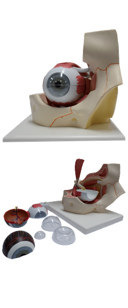
Orbit : 17 Lacrimal sac

Lacrimal Apparatus
The lacrimal apparatus consists of the:
• Lacrimal gland: secretes lacrimal fluid, a watery physiological saline containing the bacteriocidal enzyme lysozyme. The fluid moistens and lubricates the surfaces of
the conjunctiva and cornea and provides some nutrients and dissolved oxygen to the cornea; when produced in
excess, the overflowing fluid constitutes tears.
• Excretory ducts of lacrimal gland: convey lacrimal
fluid from the lacrimal glands to the conjunctival sac.
• Lacrimal canaliculi (Latin small canals): commence at a
lacrimal punctum (opening) on the lacrimal papilla
near the medial angle of the eye and drain lacrimal fluid
from the lacrimal lake (Latin lacus lacrimalis; a triangular space at the medial angle of the eye where the tears collect) to the lacrimal sac (dilated superior part of the
nasolacrimal duct).
• Nasolacrimal duct: conveys the lacrimal fluid to the inferior nasal meatus (part of the nasal cavity inferior to the
inferior nasal concha.
The lacrimal gland, almond shaped and approximately 2 cm
long, lies in the fossa for the lacrimal gland in the superolateral part of each orbit. The
gland is divided into a superior orbital and inferior palpebral parts by the lateral expansion of the tendon of the levator palpebrae superioris. Accessory lacrimal
glands may also be present, sometimes in the middle part
of the eyelid, or along the superior or inferior fornices of the
conjunctival sac. They are more numerous in the superior
eyelid than in the inferior eyelid.
Production of lacrimal fluid is stimulated by parasympathetic impulses from CN VII. It is secreted through 8-12
excretory ducts, which open into the lateral part of the superior conjunctival fornix of the conjunctival sac. The fluid
flows inferiorly within the sac under the influence of gravity.
When the cornea becomes dry, the eye blinks. The eyelids
come together in a lateral to medial sequence pushing a film
of fluid medially over the cornea, somewhat like windshield wipers. In this way, lacrimal fluid, containing foreign material such as dust is pushed toward the medial angle of the
eye, accumulating in the lacrimal lake from which it drains
by capillary action through the lacrimal puncta and lacrimal
canaliculi to the lacrimal sac.
From this sac, the fluid passes to the inferior nasal meatus
of the nasal cavity through the nasolacrimal duct. It drains
posteriorly across the floor of the nasal cavity to the nasopharynx and is eventually swallowed. In addition to cleansing
particles and irritants from the conjunctival sac, lacrimal fluid
provides the cornea with nutrients and oxygen.
The nerve supply of the lacrimal gland is both sympathetic and parasympathetic. The presynaptic
parasympathetic secretomotor fibers are conveyed from the
facial nerve by the greater petrosal nerve and then by the
nerve of the pterygoid canal to the pterygopalatine ganglion,
where they synapse with the cell body of the postsynaptic
fiber. Vasoconstrictive, postsynaptic sympathetic fibers,
brought from the superior cervical ganglion by the internal carotid plexus and deep petrosal nerve, join the parasympathetic fibers to form the nerve of the pterygoid canal and
traverse the pterygopalatine ganglion. The zygomatic nerve
(from the maxillary nerve) brings both types of fibers to the
lacrimal branch of the ophthalmic nerve, by which they enter
the gland.